


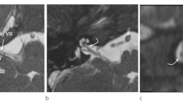
Nút phình hình túi với WEB: Thiết bị, kỹ thuật, mẹo và hiệu quả lâu dài
20/04/2021 10:50:34 | 0 binh luận
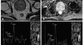
Gía trị cộng hưởng từ phổ trong chẩn đoán ung thư tuyến tiền liệt
25/03/2020 22:33:20 | 0 binh luận
T he value of magnetic resonance spectroscopy in diagnosis of prostate cancer SUMMARY Purpose: The aim of study was to determine the value of MRS in diagnosis of prostate carcinoma especially for differentiating begnin from malignant lesion of the prostate. Materials and methods : During a period of 4/2014 to 6/2016, 25 consecutive patients with elevated PSA level or clinical suspiciousness were evaluated with MRS of the prostate. The results were confirmed by TRUS-guided biopsy. We compare two groups (prostate carcinoma/PCa and prostate non-caricnoma/PNCa) by variant: mean of the choline plus creatine -to- citrate. Analyzing ROC curve to find the value of MRS in differentiating begnin from malignant tissue of the prostate. Results : Patients range in age from 40 to 89 years (mean 71 ± 12 year). 08 patients were confirmed to have PNCa (32%), whereas 17 patients had PCa (68%). The mean of (Cho+ Cr)/Ci values for PNCa and PCa were 0.50± 0.31 and 2.64± 1.22 respectively. The mean of (Cho+Cr)/ Ci value of PCa was significantly higher than PNCa (p<0.05). On ROC curve, using discrimination threshold of (Cho+Cr)/ Ci is 0.84, the MRS provided a sensitivity of 94.1%, specificity of 87.5% for differentiating NPCa from PCa. Conclusion : Magnetic Resonance Spectroscopy of the prostate can be use to differentiate begnin from malignant tissue with high accuracy. Key words: Prostatic carcinoma, non-carcinoma, Magnetic resonance spectroscopy, TRUS.
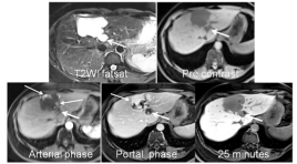
Chất tương phản MRI - PRIMOVIST: Vai trò trong chẩn đoán thương tổn gan
22/03/2020 19:18:01 | 0 binh luận
The role of hepatobiliary specific MR cantrast agent in the diagnostic focal liver lesion SUMMARY Purpose: Assessing the role of hepatobiliary - specific MR contrast Agent in the detection and differentiation of benign and malignant focal liver lesions. Maximizing accuracy of imaging in the context of focal liver lesions is paramount in avoiding unnecessary biopsies, which may result in post-procedural complications. Methods and Materials: Retrospectively evaluated the 200 patients cases, executed hepatic MRI at Medic Medical center wich hepatobiliary - specific MR contrast Agent. From on January 2013 to on January 2016, age range 28 - 72years. The lesion detected in liver: 65 cases HCC, 5 Metastasis, 2 cholangiocarcinoma, 2 Adenoma, 26 FNH, 24 Hemangiomas, 14 Regenerative nodules, 20 cysts, 5 Abscess, 37cases had normal liver. All the patients executed T2WI fatsat, Diffusion weighted imaging - MRI with three b values (0, 500, 800 sec/mm2) and Dynamic with Primovist contrast media on the Siemens Avanto 1,5T MRI. Results : In 65cases MRI HCC: 8 cases <1cm; 32 cases 1-2cm; 25 cases >25cm. 26 cases biopsic result: 24 cases HCC (92%), 2 cases cholangiocarcinoma, 39 cases TOCE or RFA no biopsy. 5 cases metastas detected primary tumor. 2 cases cholangiocarcinoma are right with biopsic result. 2 cases adenoma: 1case biopsic resulted in adenoma, 1 case follow up over 1year. 25 cases hemangiomas with contrast media from peripheral enhancement progressing to centre of lesion; 12 cases regenerative nodules is follow up over 1 year no change in nodule size; 20 cysts are high-signal intensity like fluid - signal on the T2WI, T1WI, Diffusion and no change of intensity is found after contrast injection; in 5 cases abscess: 3 cases recovered from an illness, 2 cases HCC after biopsy. Conclusion : Magnetic resonance imaging executed with hepatobiliary - specific MR contrast Agent, contributed to detect the small lesion and differential diagnosis between benign and malignant tumor of the liver.
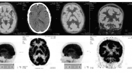
Đặc điểm hình ảnh 18F-FDG PET/CT não ở bệnh nhân Alzheimer và ở người lão hóa bình thường
25/03/2020 22:45:48 | 0 binh luận
Imaging characteristics of brain 18f-fdg pet/ct in alzheimer’s disease patients and in normal elderly persons SUMMARY Purpose : Define 18F-FDG PET/CT cerebral imaging characteristics in Alzheimer’s Disease patients and in the normal elderly persons (Nls). Objects and Methods : From 2014 to 2015, 26 Alzheimer’s disease patients and 20 normal elderly persons undergone brain 18F-FDG PET/CT scans. Results: Mean age of Alzheimer’s disease patients is 66,3±8,2 years old and 64,2±8,1 in Nls. Homogenous 18F-FDG cerebral uptake in Nls. Most of cerebral regions in Alzheimer’s disease patients suffering from a reduction of mean SUV. Rate of 18F-FDG hypometabolism in medial temporo-hippocampal area in 96.2% of cases on left side, meanwhile 92.3% of cases having a hypometabolism in right medial temporo-hippocampal area as well as in bilateral posterior cingulate gyrus, 76.9% in temporo-parietal area on right side and 86.5% on left side. Cerebral metabolism in occipital lobes principally reserved. Hypometabolism affecting bilateral frontal lobe in a half of cases. Brain glucose metabolism partially reserved at primary moto-sensory cortices, cerebellum, as well as in anterior cingulate gyrus, especially reservation of glucose metabolism at basal ganglias in Alzheimer disease (96,2%). Rate of glucose hypometabolic Alzheimer like patterns in 92,3% of cases with 84,6% on both sides. Conclusion: No focal cerebral cortical hypometabolism in Nls. Evident reduction in 18F-FDG uptake of cerebral cortices in Alzheimer’s disease patients in compared with Nls. Imaging characteristics of hypometabolism in 18F-FDG PET/CT brain scan in Alzheimer’s disease patients are regional anatomically and specifically with high rate in medial temporo-hippocampal, posterior temporal, temporo-parietal and posterior cingulate areas, well correspondent to wellknown glucose metabolic patterns in Alzheimer’s disease. Key words: Alzheimer’s Disease (AD), Brain 18F-FDG PET/ CT, imaging characteristics.
Hình ảnh cắt lớp vi tính và cộng hưởng tử bất thường dây thần inh ốc tai trên 22 bệnh nhân điếc tiếp nhận bẩm sinh
07/04/2020 21:04:15 | 0 binh luận
CT scanner and MRI imagingof cochlear nerve deficiency in 22 patients with bilateral congenital sensorineural hearing loss SUMMARY Objective: To describe CT scanner and MRI imagingof cochlear nerve deficiency (CND) and cochleovestibular nerve abnormality in association with cochlear aperture, internal auditory canal (IAC) and labyrinthine malformations. Material and Methods : 22 patients with CNDin 43 ears. Aplasia or hypoplasia of the cochlear branch was evaluated on high resolution 3D gradient-echo MRI. Cochlear aperture, IAC and bony labyrinthine malformations was evaluated on high resolutionCT scanner. Results : 22 patients with CNDin 43 ears. Cochlear nerve aplasia in 20 ears (46,5%), cochlear nerve hypoplasia in 2 ears (4,7%), presence of vestibulocochlear nerve with no cochlear branch in 21 ears (48,8%). Labyrinthine malformation in 25 ears (58,1%).The mean IAC diameter 3,03 ± 1,03mm.Cochlear aperture stenosis and atresia 76,7%. Conclusion :CND frequently associated with labyrinthine malformations, cochlear aperture stenosis or atresia and IAC stenosis. Key words : Cochlear nerve deficiency, CT scanner and MRI.
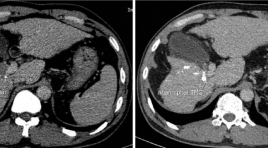
Đánh giá kết quả điều trị nút mạch hóa dầu kết hợp truyền CISPLATIN trong ung thư gan có huyết khối tĩnh mạch cửa
17/03/2020 16:39:48 | 0 binh luận
Evaluate treatment results of ctace combine with intratumoral injection of cisplatin in hepatocellular carcinoma with major portal vein thrombosis SUMMARY Advanced stage hepatocellular carcinoma with portal vein thombosis has worse prognosis and limited treatment. Transarterial chemoembolization use Lipiodol combined Cisplatin intratumoral is a safe method and have efficacy in treatment. Objective : To evaluate safety and efficay of cTACE combine with intratumoral injection of Cisplatin for hepatocellular carcinoma with major portal vein shunt treatment. Methods: From May 2018 to May 2019, 24 patients who had diagnosis of HCC with major portal vein thrombosis were perfomed cTACE (Famorubicin + Lipiodol) combined with intratumoral injection of Cisplatin. The patients were followed after treatment 1 week for the clinical symptom and after 1 month, used mRECIST and tumor markers (AFP or PIVKA-II) to aveluated treatment efficacy. The patients were perfomed consecutive courses of treatment and followed until died or until the end of study. Results: 24 patients (21 males, 3 females), age mean is 54,4 (from 32ys to 72ys, AFP mean 15600 ng/ml (from 3 to 121000), 19 patients (79%) has size of tumor ≥5cm. The classification of portal vein thrombosis: 6 patients Vp1 and Vp2; Vp3 18 patients. Total courses of treatment was 38 times. 15 patients (62,5%) had post embolization syndrome, 8 patients (33,3%) had decreasing of tumor marker. mRECIST: CR 3 patients (12,5%); PR 6 patients (25%); SD 4 patients (16,7%); PD 11 patients (45,8%). The mean survival time was 9,9 ± 1,1 months. The survival times is depend on the classification of PVTT (p=0.037 the mRECIST respondsibility (p=0.0001), the decreasing of tumor markers (p=0.01) and isn’t depend on the AFP before procedures. Conclusions : cTACE combine with intratumoral injection of Cisplatin is safety an have efficacy on treatment of HCC with major portal vein thrombosis. Keyword: Hepatocellular with portal vein thrombosis, cTACE, Cisplatin injection.
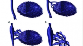
Đánh giá kết quả điều trị tiêm xơ dị dạng tĩnh mạch dưới hướng dẫn DSA
17/03/2020 16:57:22 | 0 binh luận
Evaluation the result of flouroscopy-guided sclerotherapy in treating superficial venous malformation SUMMARY Objective : Describe characteristics imaging of superficial venous malformation (VM) on flouroscopy and evaluate effectiveness of foam sclerotherapy. Methods: Prospective and retroprestive cohort from November 2015 till July 2019 on 17 patients with VM treated by flouroscopy-guided sclerotherapy with 21 lesions and 46 seasons sclerotherapy. Evualating results of treatment based on improvement in symtomps ( pain- Visual Analogue Score) and imaging ( MRI- repeat MRI after last season for 6 months . There are 4 grade in improvement in imaging: excellent ( reduction in size of lesion over 90 %), good ( reduce 50 -90 %), average ( 10-50 %) and no reponse ( less 10 %). Evualating recurrence based on increasing pain score (VAS) or size in MRI. Using SPSS 20.0 to analize and process data. Results : 17 patients (7 males and 11 females) with VM were involved in our study. Patients were a mean of 26.5± 12.9 years old ( range: from 6 to 59). Evualated by digital subtraction angiography, the lesion were categorized into 4 types according to the venous drainage features. Of the 21 lesion: 3/21 had type I (14.3 %), 12/21 had type II (57.1 %); 2/21 had type III (9.5 %) and 4/21 had type IV (19 %). Total seasons are 46. 8/21lesions (38.1 %) achieved excellent response, 9/21 (42.9 %) achieved good response, 3/21 (14.3 %) achieved average response and 1 patient (4.8%) no response in magnetic resonance imaging in magnetic resonance imaging assessments. Mean VAS scores after treatment for 1 month: 1.4; 3 months: 0.9; 6 months: 1.1, min: 0, max: 5. The long- term recurrence rates with follow-up time from 1 to 2 years is: 3/4 patients had recurrence (75 %) include of 1 patients had increasing imaging and pain score, 2 patients just had only increase imaging in MRI. Conclusion : Flouroscopy-guided sclerotherapy is a safe and effective procedure to reduce size and pain for patient with venous malformations that have symptoms. But the long – term recurrence rates is quite high. Keywords: venous malformation, sclerotherapy, flouroscopy-guided
Bạn Đọc Quan tâm
Sự kiện sắp diễn ra

THÔNG BÁO SINH HOẠT KHOA HỌC 19/01/2024: CẬP NHẬT CHẨN ĐOÁN VÀ ĐIỀU TRỊ UNG THƯ TRỰC TRÀNG
19-01-2024 09:47:01 0 bình luận

Thông tin đào tạo
- Những cạm bẫy trong CĐHA vú và vai trò của trí tuệ nhân tạo
- Hội thảo trực tuyến "Cắt lớp vi tính đếm Photon: từ lý thuyết tới thực tiễn lâm sàng”
- CHƯƠNG TRÌNH ĐÀO TẠO LIÊN TỤC VỀ HÌNH ẢNH HỌC THẦN KINH: BÀI 3: U não trong trục
- Danh sách học viên đạt chứng chỉ CME khóa học "Cập nhật RSNA 2021: Công nghệ mới trong Kỷ nguyên mới"
- Danh sách học viên đạt chứng chỉ CME khóa học "Đánh giá chức năng thất phải trên siêu âm đánh dấu mô cơ tim"


Đơn vị hợp tác

