


Nghiên cứu đặc điểm hình ảnh cộng hưởng từ sọ não trong bệnh bại não của trẻ em
07/04/2020 20:44:15 | 0 binh luận
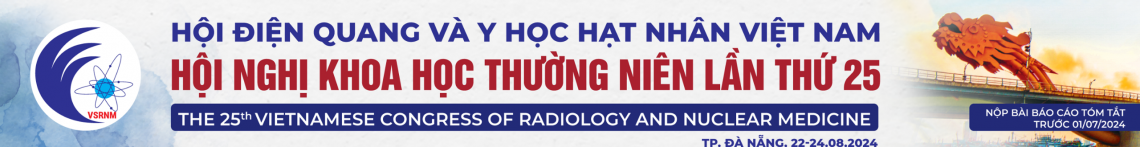
The imaging of cerebral palsy on MRI in children SUMMARY Objective : Description the image on MRI 3T of cerebral palsy and the correlation between imaging and clinical findings. Methods: From 01/2015 to 12/2016, 496 patients were diagnosed as cerebral palsy, have been done cerebral MRI in Diagnostic Imaging service. Results: 496 patients with age average 6.04 ± 4.08, youngest was under 1 year old and oldest was 15 years old. MRI shows the periventricular leukomalacia (white matter lesion) is the most popular position in CP which found in hypoxie in prenatal or perinatal, especially postnatal. The patients who had long term - hyperbilirubin have some lesion in the globus palidus and some basal ganglias. Abnormal formation of the brain, including cortical dysplasia, polymicrogyria, lissencephaly, pachygyria, schizencephaly, polymicrogyria and agenesis of the corpus callosum, have a significant rate 7.7 %. Normal MRI finding was noticed with 17.14%. Conclusion: Cerebral MRI is useful tool in confirming the degree of cerebral palsy and provides some correlation between imaging and clinical. Keywords : cerebral palsy, imaging of cerebral palsy.
Đặc điểm hình ảnh và giá trị chẩn đoán bệnh động mạch vành của xạ hình SPECT tưới máu cơ tim chụp tư thế nằm ngửa kết hợp nằm sấp
06/04/2020 22:09:43 | 0 binh luận
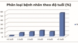
Imaging characteristics and diagnostic value of SPECT myocardial perfustion scintigraphy with combined supine - prone acquisition in diagnosis of coronary artery diseases SUMMARY Abstract: The aim of the study is to determine the imaging characteristics and diagnostic values of the combined supine - prone myocardial perfusion SPECT in comparison with supine alone acquisition. Methods : 59 CAD suspected patients were underwent Tc-99m sestamibi ECG - gated myocardial perfusion SPECT with combined supine - proneacquisition protocol to reduce attenuation defects in supine images. The results of MPI were compared with the coronary angiogram. Results: The sensitivity and specificity of the combined acquisitionprotocol were 76%, 94.1% respectively. There is no significant difference in the sensitivity between the two patient’s position acquisition. However, specificity was significantly improved in the combined acquisition (94.1% vs. 70.6%), especially in male (96.4% vs. 71.4%) and overweight patient’s groups (BMI> 23) (94.4% vs 61.1%). Conclusions : The combined supine - prone acquisitionprotocol did significantly improved the specificity ofmyocardial perfusion SPECT, especially in the overweight and males. Key words: combined supine - proneacquisition ,myocardial perfusion SPECT
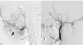
Đánh giá kết quả điều trị dị dạng tĩnh mạch nông bằng tiêm xơ dưới máy chụp mạch
06/04/2020 21:52:02 | 0 binh luận
Evaluation the result of flouroscopy-guided sclerotherapy in treating superficial venous malformation SUMMARY Purpose : Describe imaging characteristics of superficial venous malformation (VM) on flouroscopy and evaluate effectiveness of foam sclerotherapy. Method: Prospective cohort from november 2015 till may 2017 on 26 patients with VM treated by flouroscopy-guided sclerotherapy. Results: 26 patients with VM were involved in our study: 15,4% type I; 61,5% type II; 7,7% type III and 15,4% type IV. Malformation dimensionwere reduced by mean of 41,3%, VM type I had 62,5% size reduction; pain score were decreased by mean of 2,9 points (48,5%). VM type IV had 26,7% dimension reduction and 30,8% painful ease. No complication were noted. Conclusion: Flouroscopy-guided sclerotherapy is a safe procedure to reduce size and pain for patient with VM, particularly the most dimension reduction in type I, the most pain ease in type II . Keywords: venous malformation, sclerotherapy, flouroscopy-guided
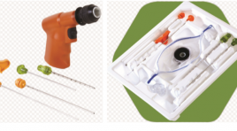
Kết quả bước đầu sinh thiết xương qua da dưới hướng dẫn cắt lớp vi tính bằng hệ thống khoan xương ARROW ONCONTROL tại bệnh viện Chợ Rẫy
03/04/2020 16:57:33 | 0 binh luận
Initial results of ct-guided percutaneous bone biopsy using an arrow oncontrol bone access system at Cho Ray Hospital SUMMARY Background: Computed tomography (CT)-guided percutaneous bone biopsy has been widely performed at big hospitals in Vietnam, but these techniques were done by using bone biopsy handle needle. Procedural time was usually prolonged, pain level of patients increased, especially osteopetrosis lesion, risk of not receiving samples. Therefore, the purpose of our study was to assess the initial effectiveness of CT-guided percutaneous bone biopsy using Arrow Oncontrol bone access system. Materials and methods: All the patients were performed CT- guided percutaneous bone biopsy at Cho Ray hospital from February 2016 to June 2017, 58 patients using Arrow Oncontrol bone access system. The techniques include: local anesthesia, confirming accurate lesion by CT scan, inserting 11G needle and 13G coaxial needle with Arrow Oncontrol bone access system putting into lesion, checking again by CT scan, drilling to take core sample. The efficacy, safety, pain level and procedural time were evaluated by variants: technique success rate, accuracy of histopathology, pain level, procedural time and complications. Results : 58 patients were performed with CT-guided percutaneous bone biopsy using drilling Arrow Oncontrol system. Technique success rate was 96.5%, diagnostic accuracy histopathology was 87.9%, the correlation was strong (r = 0.95) between histopathological diagnosis and discharge diagnosis, low pain score (mild pain) 82.7%, technical median time 11 minutes, there were no complications. Conclusions: CT-guided percutaneous bone biopsy using Arrow Oncontrol drill-assisted system had high technique success rates, high diagnostic accuracy of histopathology, mild pain level, fast and safe procedure. Key words: CT-guided bone biopsy, Arrow Oncontrol bone access system.
Vai trò của X-quang cắt lớp vi tính trong chẩn đoán lồng ruột ở người lớn
03/04/2020 16:31:14 | 0 binh luận
Role of abdominal computed tomography in diagnosis of adult intussception SUMMARY Objective: To describe imaging characteristics of adult intussception (AI) on CT and to identify diagnostic values of them. Materials and methods : Case series report including patients: defined AI on CT, over 18 years of age, having been on surgery, with/ without histo-pathology. Patients are divided into two groups: those with enteroenteric intussusception (EI) and those with intussusceptions involving colon (IC), including enterocolic and colocolic lesions. Results: from 01/2014 to 01/2017 at University Medical Center, HCMC, there were 53 intussusceptions of 52 patients on CT (EI: 14_26%, IC: 39_74%). 33 of those have intussusception on surgery (EI: 10_3%, IC: 23_70%). Mean length of intussusceptions of both groups is 7,6±4,0cm (2,4-19,6). Mean diameter of intussusceptions is 4,7±1,1cm (2,2-7). Mean interposed fat thickness is 1±0,6cm (0,1-2,6). CT and surgery characteristics of patients in EI group are of minor differences. Ratio of AI on CT with obstruction and with ischemia_necrosis are both 3,8%. Outcome of diagnosing complications by CT and by surgery are comparable. Characteristics capable of predicting presence of insstususception on surgery are: in EI group: lead point, length > 6cm, interposed fat thickness > 0,5 cm; in IC group: length > 5.65 cm, interposed fat thickness > 0,75 cm. Conclusion: Characteristics capable of predicting presence of insstususception on surgery are: lead point, length, interposed fat thickness. Key words: Computerized Tomography (CT), Intussusception, obstruction, enteric ischemia, lead point.
Điện quang can thiệp trong kiểm soát đau đầu do ung thư
17/03/2020 10:42:03 | 0 binh luận
Interventional Radiology in Cancer-related Pain Management SUMMARY Pain is a clinical symptom affecting the life quality in patients with cancer. Pain can be induced from different sources; therefore, pain management is also varied following pain mechanisms. Apart from traditional pain care therapies, interventional radiology may help in managing pain with minimal invasive techniques. This review focuses on imaging guided pain management methods used in patients with severe pain in advanced cancer stages when traditional pain control shown ineffective. Key word: Interventional Radiology, Pain management, Cancer.
Điều trị ho ra máu cấp tính bằng nút động mạch phế quản và ngoài phế quản sử dụng phối hợp 2 loại vật liệu nút mạch
01/04/2020 14:01:43 | 0 binh luận
Bronchial artery charateristics and patient outcome in patients with acute hemoptysis treated using dual embo-agents SUMMARY Objectives : This study was carried out to describe angiographic charateristics and patient outcome in patients with acute hemoptysis treated using dual embo-agents Methods: 28 patients with acute hemoptysis were included in this study. All abnormal vessels were occluded using microsphere then re-enforced with N-butyl cyanoacrylate (NBCA). Patients were followed-up one year from procedure date. Results: Technical success rate accounted for 96.4%. Almost all patients presented with bronchial artery dilatation while 46% of patients had abnormal feeding vessels arising from subclavian artery, 39% from intercostal arteries. Postprocedural complications included chest pain (4 cases, 14.3%), infection (1 patient, 3.6%), unexpected vessel occlusion (1 case, 3.6%) and bronchial artery dissection (1 subject, 3.6%). Recurrent rate calculated in one year of following up was 7.14%. Conclusion: Major angiographic abnormalities were dilatation of the bronchial artery to lung lesions and its colaterals. Dual-material embolization is an effective treatment method with significantly high rate of technial and clinical success. Keyword : Hemoptysis, bronchial artery, embolization.
Đánh giá hiệu quả nút mạch điều trị chảy máu hàm mặt do chấn thương
01/04/2020 13:55:36 | 0 binh luận
Evaluation the effectiveness of arterial embolization in the treatment of maxillofacial trauma SUMMARY Purpose: This study was designed to characterize image of vascular lesions on DSA and evaluate the effectiveness of arterial embolization in the treatment of maxillofacial trauma. Materials and Methods: 44 patients with bleeding after jaw injury, did not meet with the local hemostatic measures who were taken to the angiography for embolization from April 2011 to September 2015. Results: 13.6% of the internal carotid artery injury with 4.5% of dissection and 9.1% of carotid-cavernous sinus fistula. 90,1% of the external carotid artery injury, the (internal) maxillary artery is the most vulnerable (in 88.6%) that the maxillary artery injury merely in 56.8% or combination in 31.8%. The external carotid artery injury of a side in 59.1%, 40.9% of two side. Active bleeding is the most common of injury morphology (88,6,6%), with 63.6% merely, and associated with other forms 25%, (pseudoaneurysms, arteriovenous malformation, the internal carotid artery injury), pseudoaneurysm merely is a rare lesions in 2.3%. Hystoacryl is the most common embolization material (86.3%), 59.1% of Hystoacryl merely and coordinate 13.6%. PVA embolization merely in 13.6%; Spongel in 13.6%; no circumstances used to try Coil. Technical success was 95.4%, 4.6% failed. Successful hemostasis was achieved in 95.4% after the first intervention and 100% after 2nd intervention. Clinical success was achieved in 79.6%. Clinical non-success included 7 patients died of severe traumatic brain injury (15.9%) and 2 patients (4.5%) had complications, including 1 patient with face necrosis and 1 patient with tongue necrosis. Conclusion : Arterial Embolization in the Treatment of Maxillofacial Trauma was effective and quick to control bleeding. Keywords: bleeding, maxillofacial trauma, Embolization.
Bạn Đọc Quan tâm
Sự kiện sắp diễn ra

THÔNG BÁO SINH HOẠT KHOA HỌC 19/01/2024: CẬP NHẬT CHẨN ĐOÁN VÀ ĐIỀU TRỊ UNG THƯ TRỰC TRÀNG
19-01-2024 09:47:01 0 bình luận

Thông tin đào tạo
- Những cạm bẫy trong CĐHA vú và vai trò của trí tuệ nhân tạo
- Hội thảo trực tuyến "Cắt lớp vi tính đếm Photon: từ lý thuyết tới thực tiễn lâm sàng”
- CHƯƠNG TRÌNH ĐÀO TẠO LIÊN TỤC VỀ HÌNH ẢNH HỌC THẦN KINH: BÀI 3: U não trong trục
- Danh sách học viên đạt chứng chỉ CME khóa học "Cập nhật RSNA 2021: Công nghệ mới trong Kỷ nguyên mới"
- Danh sách học viên đạt chứng chỉ CME khóa học "Đánh giá chức năng thất phải trên siêu âm đánh dấu mô cơ tim"


Đơn vị hợp tác

