


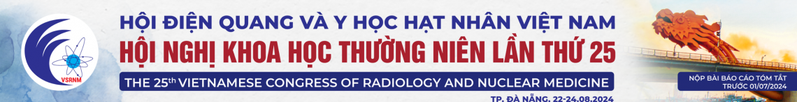
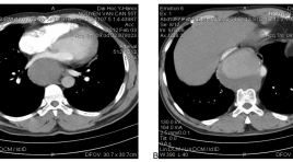
Đặc điểm hình ảnh động mạch vành sau đặt stent trên chụp cắt lớp vi tính 64 dãy
23/05/2020 11:19:07 | 0 binh luận
Imaging characteristis of coronary artery after stent implantation in 64 slice computed tomography SUMMARY Objective: Describe imaging characteristis of coronary artery after stent implantation in 64 slice computed tomography. Method: This is a prospective, cross section study applied on 85 patients who satisfied study selective requirement, from 06/2012 to 07/2013 in Hữu Nghị Hospital. Result: The most common age group is above 60 years old (90.6%); 48.2% patients have one coronary artery impairment, only 11.8% patients have three coronary arteries impairment; median diameter of stent is 3.02±0.5 mm, median length is 28.9±10.2mm; the percentage of LAD is 53.8%, RCA is 33.3%, LMA is 3.8%. There are 16 case have restenosis, in-stent lesions in 68.8% (11/16), pre-stent extending lesion in 31.2% (5/16). Conclusion: 64 slide computed tomography is non-invasive diagnosis method which have advantage in evaluate coronary artery after stent impantation: location of stent, lesions characteristic, this method also have high sensitivity and speciality in diagnoses restenosis compare to angiography. Keywords: 64 slide computed tomography, coronary artery.
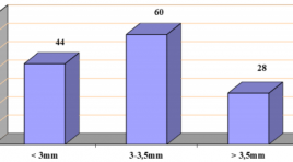
Nghiên cứu đặc điểm tổn thương trên xạ hình SPECT tưới máu cơ tim ở bệnh nhân sau nhồi máu cơ tim
02/04/2020 22:32:58 | 0 binh luận
Characteristics of myocardial perfusion on SPECT at the post - infarct affection SUMMARY: Aims: the purpose of our study was to evaluate characteristics of myocardial perfusion defects in Tc99msestamibi gated SPECT myocardial perfusion imaging (MPI). Subjects and methods: 119 post-myocardial infarction (MI) patients were underwent gated SPECT in Nuclear Medicine Department, 108 Central Military Hosspital from March 2007 to May 2010. Results: in gated SPECT MPI, reversible, mixed and fixed perfusion defects were detected in 63.9%, 18.5% and 17.6%, respectively. In patient group with ESV ≥ 70 ml, SSS and SRS were significantly higher in group with ESV < 70 ml (18.63 ± 5.02 and 15.58 ± 4.99 vs 14.49 ± 4.83 and 11.15 ± 4.63 (p<0,001). There were significant correlations between SRS and SSS with WMS (r = 0.68, p<0.001 and r = 0.61, p<0.001). In patient group with EF ≤ 40%, SSS and SRS were significantly higher in patients with ESV>40% (19.83 ± 4.36 and 17.07 ± 4.58 vs 15.50 ± 5.2 và 12.13 ± 4.85; p<0.001). There were correlation between SRS and SSS with EF (r = - 0.47, p < 0.001) và SSS (r = -0.44, p<0.001). Conclusions: In post-MI patients, fixed, reversible and mixed defects are frequently detected in SPECT MPI. The extent and severity of perfusion defects are significantly correlated with wall motion, left ventricular volume and ejection fraction evaluated by gated SPECT MPI.
Kết quả ban đầu can thiệp nội mạch trong tái thông hẹp tắc mạn tính động mạch chậu
02/04/2020 21:28:25 | 0 binh luận
The short-term primary patency of endovascular intervention in recanalrization of chronic iliac arterial occluded diseases SUMMARY: Purpose: to evaluate the short-term primary patency of endovascular intervention in recanalrization of chronic iliac arterial occluded diseases. Method and materials: prospective study, group of 21 patients with diagnosis of chronic occlusion of iliac arteries in Bach Mai hospital from 9/2011 to 6/2012. The patients were indicated to do revascularization by endovascular intervention. Result: 21 patients with 28 iliac arteries which were treated by endovascular intervention including percutaneous angioplasty alone and Stenting. Arterial access were performed at common femoral arteries bilateral (retrograde) in 100%, including one failed case that required second intervention by brachial arterial access. No complication relates arterial access. There are 89,3% procedures of stenting with angioplasty post-Stenting and 100% completed deployment of stents. No complication of angioplasty and stent deployment such as arterial rupture or perforation. The success rate of recanalrization is 96% with 96% successful rate in the first approach to cross the over the occlusion. No required 2nd reintervention in duration of follow up from 1 to 6 months, 100% of case with improved intermittent claudication and ABI. Conclusion: the initial results confirm that the endovascular intervention is a safe, effective method in the goal of recanalrization of chronic occluded iliac arteries.
Điều trị phình động mạch chủ ngực bằng stent phủ tại bệnh viện trường đại học y Hà Nội: nhân ba trường hợp
02/04/2020 20:47:46 | 0 binh luận
Thoracic endovascular aortic repair (TE VAR) for the descending thoracic aortic aneurysm at the HaNoi Medical University Hospital:report three cases SUMMARY: Thoracic aortic aneurysm is a life threatening condition that needs to be treated urgently. Thoracic endovascular aortic repair (TEVAR) for the descending thoracic aortic aneurysm was first reported by Dake et al in 1994 [4]. The advent of TEVAR was clinically relevant because descending thoracic disease repair had long been associated with operative mortality rates. The favorable results of TEVAR with reduction of operative mortality have led it to be used worldwide. Up to now, TEVAR has been initially used in Viet Nam. We report three cases with descending thoracic aorta aneurysms that were successfully treated by endovascular stent graft in HaNoi medical university hospital. Keywords: Thoracic aneurysm, stent graft, TEVAR.
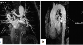
Bước đầu đánh giá vai trò của cộng hưởng từ tim trong chẩn đoán tứ chứng Fallot
02/04/2020 14:07:17 | 0 binh luận
Initial evaluation of cardiac magnetic resonance in for diagnosis of Fallot tetralogy SUMMARY: Purposes: To apply cardiac magnetic resonance (CMR) for diagnosis preoperative Tetralogy of Fallot patients and to compare the findings on echocardiography with CMR. Methods and materials: 32 patients were included in a prospective study during the interval from June 2008 to August 2009 at Bach Mai hospital. Both echocardiography and CMR had been perrformed to evaluate the variables: Ejection Fraction (EF), the right ventricular outflow tract, main pulmonary artery (MPA), left and right pulmonary artery (LPA & RPA), major aortopulmonary collateral arteries (MAPCAs)… The correlation between the findings on CMR and echocardiography was compared by using Pearson statistics. Results: 32 patients (14 males) with average age was 19.94 ± 8.20 (range: 8 -53) y. o. 100% success CMR with average time: 80.93 ± 50.41 minutes. There was a close correlation between the findings on CMR and on echocardiography with Kappa statistics r = 0.63-0.70 (p < 0.05). CMR detect more MAPCAs and additional anomalies than echocardiography. Conclusions: The results of our study indicate the usefulness of CMR for evaluation pre-operative patients with Tetralogy of Fallot. CMR has more advantages than echocardiography for detection MAPCAs and additional anomalies Key words: Tetralogy of Fallot; Cardiac magnetic resonance imaging -CMR; Echocardiography
Điểm số vôi hóa động mạch vành: tình trạng hiện tại và khuyến cáo mới 2018
17/04/2020 09:44:31 | 0 binh luận
Bệnh mạch vành(BMV) Nguyên nhân hàng đầu gây tử vong và bệnh tật tại Mỹ 50% tử vong xảy ra ở những bệnh nhân không triệu chứng. Còn một lỗ hỏng trong phát hiện BMV ở những bệnh nhân không triệu chứng . Việc tầm soát cho cả BMV không triệu chứng lâm sàng và nguy cơ phát triển BMV trên lâm sàng vẫn là 2 thách thức lớn nhất trong chăm sóc sức khỏe . Phương pháp giúp phát hiện và định lượng vôi hoá ĐM vành. Bằng chứng cho sự hiện diện của xơ vữa mạch vành Báo hiệu nguy cơ xảy ra biến cố mạch vành trong tương lai, ngoài ra sự tiến triễn của mãng vôi hoá còn chỉ ra sự tiến triễn của xơ vữa ĐMV. Giá trị tiên đoán các biến cố tim mạch và tử vong, phân tầng nguy cơ tim mạch và theo dõi điều trị

Nghiên cứu đặc điểm lâm sàng cận lâm sàng và hình ảnh chụp cắt lớp điện toán trên bệnh nhân không lỗ van động mạch phổi kèm thông liên thất
17/04/2020 09:31:14 | 0 binh luận
Không lỗ van động mạch phổi kèm thông liên thất (APSO) khá hiếm và là một dạng bệnh tim bẩm sinh phức tạp. Phức tạp là do tính đa dạng của tuần hoàn phổi [9] . Vẫn còn là một thách thức trong chẩn đoán và điều trị Khám lâm sàng, X-Quang, ECG, Siêu âm tim và DSA là những kỹ thuật kinh điển trong chẩn đoán và định hướng điều trị [9] . X-Quang phổi và ECG gợi ý chẩn đoán nhưng không đặc hiệu. Siêu âm tim ưu thế đánh giá bất thường trong tim , nhưng hạn chế trong đánh giá bất thường ngoài tim [3] DSA giúp mô tả rõ động mạch phổi và nguồn máu nuôi phổi, đo áp lực động mạch phổi và điều trị nhưng kỹ thuật mang tính xâm lấn với mức nhiễm xạ không ít. Cộng hưởng từ trong tim mạch, không xâm lấn, không nhiễm xạ, độ tương phản hình ảnh tốt nhưng hạn chế nhiều ở khâu thực hiện Chụp cắt lớp vi tính có thể đánh giá các bất thường trong tim và tỏ ra ưu thế trong đánh giá bất thường ngoài tim: Đo kích thước động mạch phổi. Ống động mạch vị trí bất thường. Tuần hoàn bàng hệ chủ phổi. Bất thường động mạch vành. Hạn chế do nhiễm xạ. Xác định tuần hoàn phổi trong bệnh lý APSO có thể nói là khâu then chốt để đưa ra chiến lược điều trị. Chỉ định chụp MSCT để khảo sát bệnh lý này, mới và còn nhiều quan điểm khác nhau nên chúng tôi tiến hành đề tài này với các mục tiêu sau: 1- Mô tả đặc điểm lâm sàng. 2- Mô tả đặc điểm huyết học, ECG, X-Quang. 3- Khảo sát đặc điểm hình ảnh siêu âm tim và hình ảnh MSCT trên bệnh nhân APSO. 4- Khảo sát sự phù hợp giữa siêu âm tim và hình ảnh chụp cắt lớp điện toán.
Hình ảnh bệnh cơ tim phì đại (Imaging of Hypertrophic Cardiomyopathy)
16/04/2020 23:46:28 | 0 binh luận
Nội dung: Giới thiệu Các kiểu hình bệnh cơ tim phì đại (BCTPĐ) Các yếu tố nguy cơ Chiến lược chẩn đoán Điều trị và theo dõi Kết luận
Bạn Đọc Quan tâm
Sự kiện sắp diễn ra

THÔNG BÁO SINH HOẠT KHOA HỌC 19/01/2024: CẬP NHẬT CHẨN ĐOÁN VÀ ĐIỀU TRỊ UNG THƯ TRỰC TRÀNG
19-01-2024 09:47:01 0 bình luận

Thông tin đào tạo
- Những cạm bẫy trong CĐHA vú và vai trò của trí tuệ nhân tạo
- Hội thảo trực tuyến "Cắt lớp vi tính đếm Photon: từ lý thuyết tới thực tiễn lâm sàng”
- CHƯƠNG TRÌNH ĐÀO TẠO LIÊN TỤC VỀ HÌNH ẢNH HỌC THẦN KINH: BÀI 3: U não trong trục
- Danh sách học viên đạt chứng chỉ CME khóa học "Cập nhật RSNA 2021: Công nghệ mới trong Kỷ nguyên mới"
- Danh sách học viên đạt chứng chỉ CME khóa học "Đánh giá chức năng thất phải trên siêu âm đánh dấu mô cơ tim"


Đơn vị hợp tác

