


TƯƠNG QUAN GIỮA ĐƠN VỊ HOUNSFIELD Ở CỘT SỐNG THẮT LƯNG VÀ MẬT ĐỘ XƯƠNG ĐO BẰNG DEXA Ở NGƯỜI VIỆT NAM
17/10/2023 17:41:40 | 0 binh luận
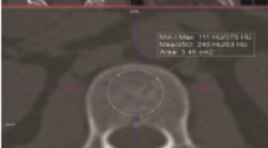
SUMMARY Background: Osteoporosis is a disease that increases the risk of fractures. Evaluation of the Hounsfield Unit in the lumbar spine on computed tomography for any reason has the potential to predict bone density abnormalities that contribute to the warning of osteoporosis. Objective: The aim of this study was to evaluate the correlation between Hounsfield Units (HU) in Lumbar Spine (LS) and bone mineral density (BMD) measured by Dual-Energy X-Ray Absorptiometry (DEXA). Method: Retrospective and cross-sectional study of 150 patients comprised CT-DXA pairs within a 6-month period performed for any indication. Measure HU at lumbar vertebrae from L1 to L4. Calculate Spearman correlation coeffificient between the mean HU at lumbar vertebrae and the BMD values from DEXA scan. Using area under the ROC Curve (AUC) and finding cut-off of HU for diferentiating normal BMD and abnormal BMD, osteopenia and osteoporosis. Result: We noted correlations between the HU at LS and the BMD from DXA scan which is significant, the highest correlation at L2 (Spearman correlation coefficient = 0.68). At L2, normal BMD: ≥ 131 HU, osteopenia: 101 – 131 HU, osteoporosis: ≤ 131 HU, we also determined that threshold of 101 HU was more than 90 % sensitive, and a threshold of 171 HU was more than 90 % specific for distinguishing normal BMD. In addition, cut-off ≤ 132 HU was more than 90 % sensitive, and cut-off ≤ 62 HU was more than 90 % specific for distinguishing osteoporosis from osteopenia. Conclusion: The correlations between the HU at LS and the BMD from DXA scan is significant. Keyword : Osteoporosis, DEXA, HU at lumbar spine
NGHIÊN CỨU ĐẶC ĐIỂM TỔN THƯƠNG XƯƠNG TRÊN XẠ HÌNH Ở BỆNH NHÂN UNG THƯ PHỔI KHÔNG TẾ BÀO NHỎ GIAI ĐOẠN IIB - IVB TẠI BỆNH VIỆN UNG BƯỚU THÀNH PHỐ CẦN THƠ NĂM 2020 - 2021
16/10/2023 16:48:04 | 0 binh luận
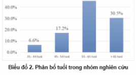
SUMMARY Background: Non-small cell lung cancer (NSCLC) is the most common cancer and has a high rate of bone metastases. Early detection of bone metastases is important in treatment and improves the quality of life for. Currently, there have been many studies on the value of bone scan in early detection of bone metastases, but this issue has not been fully evaluated. Objective: To evaluate the characteristics of bone metastases on scintigraphy in lung cancer. Methods: Cross-sectional description of a series of diseases, retrospective data on 151 patients with stage IIB - IVB non-small cell lung cancer, examined by bone scan with Tc-99m MDP (Methylene diphosphonate) before specialized treatment during the period from January 2020 to December 2021 at Can Tho City Oncology Hospital. Results: The incidence of men and women were 62,9% and 37,1%. The mean age is 60, the youngest is 35 and the oldest is 83. There are 56 patients (37,1%) with bone lesions on the scan. There are 47 lesions (83,9%) could be known as bone metastases: 46 cases are at stage IV and the other is at stage IIIB. The most common sites of lesions are from the ribs and sternum. Others are from thoracic spine, the sacrum and the coccyx. The bone lesions from the clavicle and upper extremities are rare. Most of bone lesions are multifocal, asymmetrical, and strongly radioabsorbed. Conclusion: Bone-image characteristics from scan in stage IIB – IVB non-small cell lung cancer have a high incidence of bone metastases. Therefore, the application of bone scan as a routine subclinical in the initial diagnosis of NSCLC before treatment is essential in order to properly assess the stage of the disease, make an accurate prognosis and have a reasonable treatment strategy. Keywords: Bone scan, non-small cell lung cancer
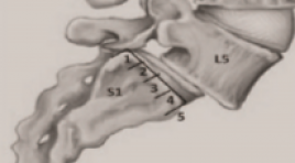
ĐÁNH GIÁ SỰ CẢI THIỆN MỨC ĐỘ TRƯỢT THÂN ĐỐT SỐNG SAU PHẪU THUẬT TLIF DỰA TRÊN X-QUANG THƯỜNG QUY
16/10/2023 16:25:53 | 0 binh luận
SUMMARY Aim: The study was carried out with the aim of evaluating the improvement of degree of vertebral body slippage after spinal fixation posterior slide manipulation with disc removal, by combining vertebaral body fusion TLIF surgery and assessingthe improvement in the level of post-X-ray-based vertebral slip, with clinical comparison. Method: A total of 39 patients diagnosed with lumbar vertebral stem slide who were surgically treated at the Department of Orthopedic and Spinal Injuries of Bach Mai Hospital between July 2021 and July 2022 were included in the study. Results: The results of the study showed that patients with lumbar vertebral slippage experienced the most in the L4-L5 position accounting for 67.4%, followed by L3-L4 accounting for 22.5%, L5-S1 accounting for 13.1%. All 100% of patients improved in the height of the disc gap between the vertebrae. No more diseases showed signs of ladder. Signs of intermittent pain also improved, and 80 percent of patients were able to travel longer distances than before. Conclusion: Postoperative X-ray examination showed that thepatients were corrected in surgery quite well, all patients had reduced levels of postoperative slippage and increase intervertebral space height. Keywords: routine X-ray, lumbar vertebral stem slide, TLIF surgery
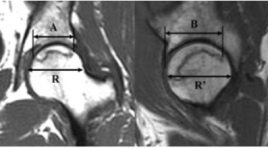
GIÁ TRỊ CỦA CỘNG HƯỞNG TỪ TỐI THIỂU TRONG CHẨN ĐOÁN HOẠI TỬ VÔ KHUẨN CHỎM XƯƠNG ĐÙI GIAI ĐOẠN SỚM Ở NHỮNG BỆNH NHÂN CÓ YẾU TỐ NGUY CƠ
11/10/2023 12:20:14 | 0 binh luận
Summary Purpose: Evaluate the agreement between limited MRI, which using T1W sequence or STIR sequence in coronal direction, with standard MRI in diagnosis early femoral head necrosis(FHN) occurring in high risk patients. Subjects and methods : descriptive cross-sectional study was performed on 58 patients, who warediagnosed of femoral head osteonecrosis at stage 2 or higher according to the Arlet Ficat classification. The patients were performed hip joints MRI at the Radiology Center, Bach Mai Hospital from June 2020 to August 2021. Results: The agreement in FHN staggingbetween limited MRI using T1Wsequence, or limited MRI using STIR sequence with the standard MRI was 0.98 and 0.86, respectively. The agreement in measurement extent of osteonecrosis areabetween limited MRI using T1Wsequence, or limited MRI using STIR sequence with the standard MRI was 0.98 and 0.85. Conclusion : There was excellent agreement between the full and limited MR examinations both forstagging and determining the extent of osteonecrosis area. The time and potential cost reduction achieved when taking limited MRImay lead to more widespread using in patient care. Key words: femoral head osteonecrosis, hip MRI, limited hip MRI.
Chẻ đôi đốt sống thể kín: Báo cáo loạt ca
30/03/2020 22:50:48 | 0 binh luận
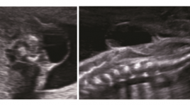
Closed spina bifida: Case series report ABSTRACT: Introduction : Imaging of closed spina bifida is very multiform, can diagnose in utero. Objective: Describe the imaging features of closed spinal bifida, classify the usual closed spina bifida. Methods: case series report, prospective study Conclusions: The important role of ultrasound in prenatal closed spinal bifida diagnosis. Identify the cranial sign and spinal defection to classify the type of closed spina bifida, to make fetal prognosis , to orientate in antenatal counseling Key words: Closed spina bifida, Spinal dysphaphism
U tế bào quanh mạch nguyên phát ở xương chày - Báo cáo một trường hợp hiếm và tổng kết trên y văn
26/03/2020 22:33:38 | 0 binh luận
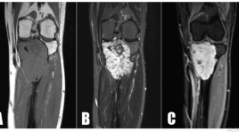
Primary haemangiopericytomas of tibia - a rare case report and review in litterature SUMMARY Hemangiopericytomas (HPCs) are rare vascular tumors arising from pericytes. Therefore HPCs have a wide distribution in both soft tissue and skeletal system, with the latter being the most unusual occurrence. Currently, there are about 74 cases of osseous HPC have been reported in the available English literature, only five of them located in the tibia. Primary hemangiopericytomas of bone usually occurs in pelvis, vertebrae and long bones of lower extremities. The prognosis of the tumor behaviour is still not feasible and it has the potential to demonstrate a highly malignant course. We introduce a case of primary HPCs of bone located in tibia, which received a treatment by widen resection and bone transportation. Keywords: u tế bào quanh mạch, u xương/ hemangiopericytoma, bone tumor.

Giá trị sinh thiết xương chi qua da dưới hướng dẫn cắt lớp vi tính trong chẩn đoán u xương
21/11/2019 16:42:05 | 0 binh luận
Sinh thiết tủy xương và chọc hút tủy xương là các thủ thuật lấy mẫu và khảo sát tủy xương –mô xốp bên trong các xương lớn. Tủy là nơi sản sinh tế bào hồng cầu, bạch cầu và tiểu cầu từ các tế bào gọi là “tế bào gốc”.

Chẩn đoán hình ảnh bệnh lý nhiễm trùng cột sống
21/11/2019 15:33:46 | 0 binh luận
Viêm cột sống nhiễm trùng chiếm 2-4% các nhiễm trùng xương. Nhiễm trùng cột sống có thể lan theo đường máu từ các ổ nhiễm trùng ở xa, vấy nhiễm trực tiếp từ phẫu thuật cột sống hoặc vết thương thấu hoặc lan trực tiếp từ các ổ nhiễm trùng mô mềm kế cận. Các tác nhân nhiễm trùng xuất phát từ máu có thể đến cột sống theo dòng tới qua các tiểu động mạch nuôi của thân sống hoặc theo dòng lui qua đám rối tĩnh mạch
Bạn Đọc Quan tâm
Sự kiện sắp diễn ra

THÔNG BÁO SINH HOẠT KHOA HỌC 19/01/2024: CẬP NHẬT CHẨN ĐOÁN VÀ ĐIỀU TRỊ UNG THƯ TRỰC TRÀNG
19-01-2024 09:47:01 0 bình luận

Thông tin đào tạo
- Những cạm bẫy trong CĐHA vú và vai trò của trí tuệ nhân tạo
- Hội thảo trực tuyến "Cắt lớp vi tính đếm Photon: từ lý thuyết tới thực tiễn lâm sàng”
- CHƯƠNG TRÌNH ĐÀO TẠO LIÊN TỤC VỀ HÌNH ẢNH HỌC THẦN KINH: BÀI 3: U não trong trục
- Danh sách học viên đạt chứng chỉ CME khóa học "Cập nhật RSNA 2021: Công nghệ mới trong Kỷ nguyên mới"
- Danh sách học viên đạt chứng chỉ CME khóa học "Đánh giá chức năng thất phải trên siêu âm đánh dấu mô cơ tim"


Đơn vị hợp tác

