


BÁO CÁO CA LÂM SÀNG: ĐIỀU TRỊ TÚI GIẢ PHÌNH ĐỘNG MẠCH VỊ TÁ TRÀNG DỌA VỠ BẰNG PHƯƠNG PHÁP CAN THIỆP NỘI MẠCH
11/10/2023 15:28:24 | 0 binh luận
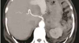
SUMMARY Gastroduodenal artery (GDA) pseudoaneurysm is a rare surgical entity that causes various symptoms. In the case of rupture, it usually presents an ominous prognosis and mortality rate of up to 40%. Although open surgical procedure is a mainstay, endovascular intervention is emerging a promising treatment in recent years, due to its advantages and safety.We present a case of upper gastrointestinal bleeding caused by GDA pseudoaneurysm threatened rupture in a 71-year-old woman, with medical episodes of acute pancreatitis, a pancreatic body tumor removal surgery was performed, and now the tumor is relapsing and metastasizing. The treatment approach is blocking off the pseudoaneurysm by a covered stent. The procedure was successful and the patient is asymptomatic. Two months later, the pseudoaneurysm reduces its size and completely excluded from the preservation of the blood flow in the artery. Endovascular interventional treatment in the case of GDA aneurysms is considered a promising alternative not only to open surgery but also to an effective emerging technique even in the acute setting. Keywords : Gastroduodenal artery, Covered stent, Pseudoaneurysm
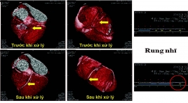
GIÁ TRỊ CỦA CẮT LỚP VI TÍNH 256 DÃY TRONG CHẨN ĐOÁN BỆNH ĐỘNG MẠCH VÀNH Ở BỆNH NHÂN RUNG NHĨ
11/10/2023 15:05:26 | 0 binh luận
SUMMARY Objectives: To determine the diagnostic value of 256 MDCT in the diagnosis of coronary artery disease in patients with atrial fibrillation. Materials and methods: We prospectively enrolled 48 patients with atrial fibrillation who underwent 256 MDCT coronary CT angiography scan at Huu Nghi Hospital from 07/2020 to 07/2021 and evaluated diagnosis value of this technique using invasive coronary angiography as golden standard. Results: At patient-based analysis, sensitivity, specificity, positive predictive value and accuracy are 100%, 83.3% and 83.3%, respectively. At vessel-based analysis, sensitivity, specificity, positive predictive value, negative predictive value and accuracy are 98.4%, 81.7%, 80.3%, 98.5% and 88.9%, respectively. At segment-based analysis sensitivity, specificity, positive predictive value, negative predictive value and accuracy are 96.7%, 93.8%, 78.4%, 99.2 % and 94.4%, respectively. Conclusion: 256 MDCT coronary angiography scan is a diagnostic method with high accuracy in diagnosing coronary artery disease in patients with atrial fibrillation. Keywords: atrial fibrillation, coronary CTA, diagnostic value
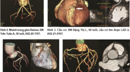
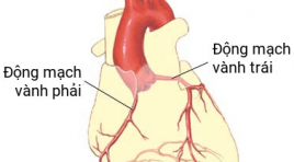
NHẬN XÉT MỘT SỐ BIẾN THỂ VÀ BẤT THƯỜNG ĐỘNG MẠCH VÀNH TRÊN CẮT LỚP VI TÍNH 128 DÃY
11/10/2023 12:17:03 | 0 binh luận
Summary Purpose: To describe the variants and the anormalies of coronary artery on 128-row computed tomography (CT). Material and method : 90 patients were treated at 103 hospital from Oct. 2020 to Jul. 2021. The characteristic figures of coronary artery assessed on 128-row CT included: the site of ostium, course, abnormal enlargement of the branch… Results : 84.4% of the patients had a right dominant system and Ramus intermedius branch was seen in 57.8%. The anomalous origins of LM and RCA were found in 3.3% for each branch. The atrophies of LCx and RCA were 7.8% and 2.2%, respectively. Conclusion : 128-row detector CT images is helpful for detecting the variants and the anormalies of coronary artery. Key words: computed tomography, coronary artery, variant, anormaly.
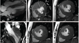
Khảo sát đặc điểm hình ảnh bắt thuốc thì muộn trên cộng hưởng từ của bệnh cơ tim phì đại
06/05/2021 11:33:36 | 0 binh luận
SUMMARY Objective: To describe the characteristics of hypertrophic cardiomyopathy on Magnetic resonance imaging; to evaluate the loacation, pattern and extent of late gadolinium enhancement (LGE); to evaluate the relationship among left ventricular function, end diastolic wall thickness and LGE about pattern and extent. Materials and Methods: Cine imaging and delayed enhancement imaging were performed in 27 patients with HCM on a 3 Tesla MRI unit (Siemens Verio) at the University Medical Center Hospital between January 2016 and June 2020. Global left ventricular function was quantified, using a Argus function software of Siemens Healthineers. The location, pattern, and extent of DE were evaluated. Results: Global left ventricular function and mass calculations yielded a mean ± SD for ejection fraction of 64.8 ± 11.7%, an end-diastolic volume of 111.5 ±27.2ml, and a left ventricular mass of 181.4 ± 96.2g. Diffuse hypertrophy was present in 12 patients (44.5%), asymmetric septal hypertrophy in 11 patients (40.7%), and apical hypertrophy in 4 patients (14.8%). LGE occurred in 24 patients (88.9%) and in 164 segments (33.7%), most commonly in the anteroseptal and inferoseptal segments. LGE was detected in an ill-defined patchy pattern in 61.6% and in a focal nodular pattern in 38.4% enhanced segments. LGE with an extent ≥ 50% was observed in 61 segments (37.2%), and that with an extent < 50% was observed in 103 segments (62.8%). There were significant difference in EF between the LGE-positive patients and the LGE-negative patients (p = 0.03). The myocardial wall was thicker in the enhanced segments than in the non-enhanced segments (p < 0.001). No significant difference was found in wall thicker of segments between ill-defined patchy pattern and focal nodular pattern in our study. The enhanced segments with the transmural extent ≥ 50% were thicker than non-enhanced segments and the enhanced segments with an extent < 50% were thicker than nonenhanced segments at end-diastole and at end-systole (p < 0.01). Conclusion: Cardiac MR imaging is beneficial in making a diagnosis and determining the phenotype of HCM because it can observe the cardiac morphology clearly and evaluate its function comprehensively. It is possible to accurately measure the wall thickness, detect high-risk phenotypes and determine myocardial fibrosis based on late myocardial enhancement. Therefore, it is necessary to perform cardiac MR imaging in patients with HCM or suspected HCM on clinical examination. Keywords: Hypertrophic cardiomyopathy, late gadolinium enhancement
Cập nhật chụp cắt lớp vi tính trong bệnh lý tim mạch
17/03/2020 10:59:11 | 0 binh luận
CT scan of cardiovascular disease: An update SUMMARY Coronary artery disease is the leading cause of death in the United States, in developing countries, coronary artery disease is rising rapidly. It’s been projected that by 2020, chronic diseases will account for almost three-fourths all deaths, 71% deaths due to ischemic heart disease. Early detection of coronary artery lesion helps to reduce mortality and improve quality of life by risk factors control and stabilize atherosclerotic plaques. The newest commercially available MDCT scanners offer the greatest degree of spatial and temporal resolution owing to multidetector channels, fast gantry rotation times and thinner collimation. These parameters are important to creation of high definition coronary artery MDCT imaging. In the case of acute chest pain, CT also proved safe, effective, low-cost, reduced the number of emergency visits and hospitalization. In addition to the accurate diagnosis, CT of coronary artery disease also evaluated other heart diseases such as congenital heart disease, pericardium, cardiomyopathy, heart valve New developments such as FFR-CT, iFR-CT or Myocardial perfusion assessment show that computed tomography for cardiovascular disease diagnosis will be more potential in the future. Keywords: coronary computed tomographic angiography, cardiac computed tomography, multi-detector computed tomography for cardiovascular disease diagnosis.
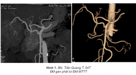
Nghiên cứu các biến đổi giải phẫu của động mạch thân tạng và mạc treo tràng trên trên chụp cắt lớp vi tính 64 dãy
23/05/2020 11:24:06 | 0 binh luận
SUMMARY The purpose of this study was to determine the different variants of celiac and superior mesenteric arteries identified at abdominal 64-slice CT scanner performed in 910 patients. Results : 282 patients (30.9%) had arterial variants identified. The most common variant identified was a right hepatic artery originating from the superior mesenteric artery, which was seen in 77 (8.5%) patients. The second most common variant was a left hepatic artery originating from the left gastric artery, seen in 47 (5.2%) patients. Variations in the origin of the common hepatic artery were seen in 10 (1.1%) patients. The right hepatic artery originated from the celiac axis in 13 (1.4%) patients and from the aorta in 3 (0.3%) patient. The left hepatic artery originated from the celiac axis in 4 (0.4%) patient. Accessory hepatic arteries were identified in 53 (5.8%) patients.
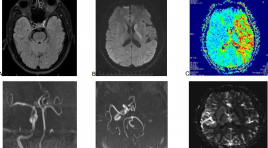
Kết quả ban đầu điều trị nhồi máu não tối cấp bằng dụng cụ lấy huyết khối Solitaire kết hợp tiêu sợi huyết đường động mach: nhân 2 trường hợp
02/04/2020 21:17:47 | 0 binh luận
Primary results of treatment of hyper acute ischemia by thrombus retrieve with Solitaire devices associated with intra-arterial fibrolytic with rtPA: Report of 2 cases SUMMARY: Brain ischemia accounts for 85% of brain stroke and this is 3rd leading cause of mortality or mobility in the world. The treatment of hyper acute ischemia by intravenous infusion of fibrolytic (rtPA) has been recently progressive. However, this indication is only selected for the patient coming early during first 3 hours after onset and it also gets some limitations in cases of main artery occlusion. The super selective endovascular treatment of brain ischemia by using thrombus retrieve device (solitaire) associated with fibrolytic (rtPA) has been confirmed to be increased the rate of recanalization and good outcome recently. We want to report our 2 cases of treatment of hyper acute ischemia by clot retrieve with Solitaire devices and fibrolytic with rtPA and review the literature.
Chẩn đoán hình ảnh ù tai theo nhịp mạch
04/01/2020 15:28:58 | 0 binh luận
Chứng ù tai theo nhịp mạch không giống như chứng ù tai vô căn, thường có một nguyên nhân cụ thể và có thể xác định được.
Bạn Đọc Quan tâm
- Ca lâm sàng: điều trị ung thư tuyến giáp bằng I-131
- Những tiến bộ trong chẩn đoán hình ảnh bệnh tim – mạch
- Ứng dụng CNTT trong chẩn đoán hình ảnh giúp tăng chất lượng y tế
- CA LÂM SÀNG: UNG THƯ BIỂU MÔ NỘI ỐNG TUYẾN VÚ
- KẾT QUẢ ỨNG DỤNG XẠ HÌNH THẬN ĐỘNG TẠI BỆNH VIỆN NHI ĐỒNG THÀNH PHỐ
- NGHIÊN CỨU BƯỚC ĐẦU ĐÁNH GIÁ VAI TRÒ CỦA TRÍ TUỆ NHÂN TẠO TRONG PHÂN TÍCH KẾT QUẢ X QUANG NGỰC THẲNG TẠI BỆNH VIỆN CHỢ RẪY
- NGHIÊN CỨU ĐẶC ĐIỂM HÌNH ẢNH VÀ VAI TRÒ CỦA SIÊU ÂM TRONG CHẨN ĐOÁN U TUYẾN CẬN GIÁP
- ĐÁNH GIÁ KẾT QUẢ BƯỚC ĐẦU ĐIỀU TRỊ TĂNG SẢN LÀNH TÍNH TUYẾN TIỀN LIỆT CÓ BÍ TIỂU CẤP BẰNG PHƯƠNG PHÁP CAN THIỆP NÚT ĐỘNG MẠCH TUYẾN TIỀN LIỆT
- VỠ TÚI GIẢ PHÌNH KHỔNG LỒ TÂM THẤT TRÁI: BÁO CÁO 1 TRƯỜNG HỢP HIẾM GẶP TẠI BỆNH VIỆN BẠCH MAI
- VAI TRÒ CỦA SINH THIẾT BẰNG KIM LÕI DƯỚI HƯỚNG DẪN SIÊU ÂM TRONG ĐÁNH GIÁ HẠCH CỔ BẤT THƯỜNG
Sự kiện sắp diễn ra

THÔNG BÁO SINH HOẠT KHOA HỌC 19/01/2024: CẬP NHẬT CHẨN ĐOÁN VÀ ĐIỀU TRỊ UNG THƯ TRỰC TRÀNG
19-01-2024 09:47:01 0 bình luận

Thông tin đào tạo
- Những cạm bẫy trong CĐHA vú và vai trò của trí tuệ nhân tạo
- Hội thảo trực tuyến "Cắt lớp vi tính đếm Photon: từ lý thuyết tới thực tiễn lâm sàng”
- CHƯƠNG TRÌNH ĐÀO TẠO LIÊN TỤC VỀ HÌNH ẢNH HỌC THẦN KINH: BÀI 3: U não trong trục
- Danh sách học viên đạt chứng chỉ CME khóa học "Cập nhật RSNA 2021: Công nghệ mới trong Kỷ nguyên mới"
- Danh sách học viên đạt chứng chỉ CME khóa học "Đánh giá chức năng thất phải trên siêu âm đánh dấu mô cơ tim"


Đơn vị hợp tác

