


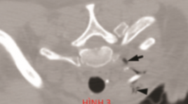
KHẢO SÁT VỊ TRÍ LỖ THÔNG XOANG BƯỚM Ở NGƯỜI TRƯỞNG THÀNH TRÊN CẮT LỚP VI TÍNH
17/10/2023 15:27:45 | 0 binh luận
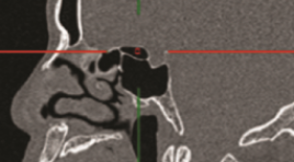
SUMMARY Background: The sphenoid sinus is closely surrounded by many important vascular and neurological. The natural ostium is the safest place to enter the sphenoid sinus without injuring adjacent structures. A better understanding of the position and distance of the sphenoid ostium (SO) with respect to the other anatomic landmark is essential for the safety and effectiveness of laparoscopic surgery. Objective: The purpose of this study was to investigate the relationship between the SO and surrounding landmark structures. Besides, we examine the effect of the Onodi cell and pneumatization of the sphenoidal sinus on the position of the SO. Methods: Cross-sectional study of 162 sinus CT scan data obtained from the PACS system at University Medical Center Ho Chi Minh City. The image is reconstructed and measured by the Multiplanar reconstruction (MPR) software of the PACS Carestream. Results: The mean distance between the SO and the lateral wall was 9.1 ± 1.8 mm. The mean distance from the SO to the median line was 4.1 ± 1.8 mm. The mean distance between the SO and the roof sphenoid sinus was 7.9 ± 2.9 mm. The average distance from the sphenoid ostium to superior border of posterior choana was 12.9 ± 3.5 mm. The mean distance from the sphenoid ostium to the anterior nasal spine was 65.2 ± 4.5 mm and to the posterior wall of the sphenoid sinus was 12.8 ± 2.7 mm. The angle between the SO - the anterior nasal spine and the floor of the nose is 33.5 ± 3.5 degrees. The distance from the SO to the roof sphenoid sinus was found to increase in cases where Onodi cells are present. For lateral pneumatization, the distance from the SO to the lateral wall in type II to be less in type III. The sphenoid sinus pneumatization on the sagittal plane did not affect the distance from SO to the posterior choana and the roof sphenoid sinus. Conclusions: The study determined the distances between the SO and some surrounding anatomical landmarks, and also investigated the influence of the Onodi cell and pneumatization on the position of the sphenoid sinus by CT scan images. The measurements described in this study may be very valuable in avoiding serious complications while performing surgery. Keywords: Sphenoid ostium, computed tomography.
XÁC ĐỊNH TỔN THƯƠNG ĐƯỜNG DẪN TRUYỀN GIỮA HAI BÁN CẦU ĐẠI NÃO SAU CHẤN THƯƠNG ĐẦU BẰNG HÌNH ẢNH HỌC MRI - DTI (DTI: DIFFUSION TENSOR IMAGING): BÁO CÁO CA LÂM SÀNG
16/10/2023 17:13:10 | 0 binh luận
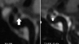
SUMMARY Traumatic brain injury (TBI) is a form of acquired brain injury resulting from an external mechanical force to the head make up the alternation of brain function. However, it is difficult to diagnose the injury to the neural tract and the connection between us through traditional imaging techniques. A 54y woman came to our clinic because of insufficient coordination of her body. Her personal history: Severe trauma brain injury with coma in 10 days treated by medical treatment 10 years ago. Some bad conditions after TBI include impairment of memory, insufficiency of coordination of her body, a post-concussion syndrome was suggested. Clinical examination: GCS: 15, strength testing: 5/5 for both sides but our patient can’t walk. She was examined at many medical centers but no evidence conforming her symptom. Our patient scanned by MRI ((Magnetic resonance imaging) Siemens 3.0 Tesla Spectra system. MRI morphometry used for her detected a significant decline of corpus callosum. MRI - DTI (Diffusion Tensor Imaging) revealed a decreased FA in the white matter of the right temporal and corpus callosum. Fractional anisotropy is a scalar value between zero and one that describes the degree of anisotropy of a diffusion process. A decrease in the value of FA in corpus callosum is indicative of the loss of connection between both hemispheres. MRI tractography used to describe the amount of neural tracts in corpus callosum. So, MRI-DTI and MRI Tractography served as a powerful diagnostic tool, providing imaging results that offered an explanation for our patient’s clinical picture Keyword: Trauma brain injury, Cerebral atrophy, Diffusion tensor imaging, Tractography, Morphometry
ĐẶC ĐIỂM HÌNH ẢNH PHÌNH ĐỘNG MẠCH NÃO TRÊN CỘNG HƯỞNG TỪ 3 TESLA
13/10/2023 18:04:10 | 0 binh luận
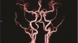
SUMMARY Objective: Describe the imaging characteristics of an intracranial aneurysm on 3 Tesla magnetic resonance at Bach Mai hospital Methods : A prospective study was performed on 29 patients with clinical manifestations of neuropathy who were diagnosed with 3 Tesla magnetic resonance brain aneurysms and then underwent digital subtraction angiography at the Radiology Center, Bach Mai Hospital, Hanoi, Vietnam from August 2021 to August 2022. Description of intracranial aneurysms in terms of number, location, shape, morphology and size measure. Results: Of the 29 patients, 37 aneurysms were detected on 3 tesla magnetic resonance and confirmed on digital subtraction angiography. The location of intracranial aneurysms is common in the anterior circulation (97.29%), and the posterior circulation (2.71%). Dimensions are mainly ≤ 10mm (97.3%). Usually saccular aneurysms (94.6%) and narrow neck (51.43%). Most aneurysms have irregular margins, knobs or two bases (72.97%). Conclusion : Magnetic resonance 3 Tesla can replace digital subtraction angiography erasing the background to characterize the image of intracranial aneurysms. This is a safe, non-invasive and highly valuable imaging method in the diagnosis of intracranial aneurysms, a very effective first choice for screening intracranial aneurysms and providing information for neurologists implement appropriate treatment strategies. Keywords: 3T magnetic resonance, intracranial aneurysm
ĐẶC ĐIỂM HÌNH ẢNH CLVT ĐA DÃY VỠ XƯƠNG HÀM MẶT DO TAI NẠN GIAO THÔNG
13/10/2023 16:55:55 | 0 binh luận
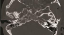
SUMMARY Objectives : study the multi-slice CT (MSCT) imaging in the diagnosis of maxillofacial fractures by traffic accident. Subjects and methods: A cross-sectional descriptive study of 63 patients with maxillofacial trauma due to traffic accidents who underwent MSCT scan at Viet Duc Hospital in April 2022. Results: 46 male and 17 female. The mean age was 28.4±12.6 (from 15 to 75 years old). The most common traffic accidents are motorbikemotorcycle accidents with 21/63 (33.3%), self-motorbike-riding accidents are 18/63 (28.6%), car-motorcycle accidents account for 16 /63 (25.4%), pedestrian - car/motorcycle accidents are 5/63 (7.9%) and car-car accidents are 3/63 (4.8%). There were 28/63 (44.4%) cases associated with cranial fracture and 32/63 (50.8%) skull base fracture. In cases of maxillofacial fracture, orbital wall fracture was the most common with 41/63 (65.1%), maxillary fracture 39/63 (61.9%), zygomatic fracture 32/63 (50.8%), nose bone 19/63 (30.2%), frontal sinus wall 13/63 (20.6%), palatal and mandibular fracture were 11/6 (17.5%). The most common type of maxillary fractures is Lefort 1 with 11/63 (17, 5%) on both right and left side, Lefort 2 and 3 fractures are less common with 1.6% to 6.3%, respectively. Conclusion : MSCT is a reliable method in diagnosing maxillofacial fractures. Keywords: maxillofacial trauma, traffic accident, computer tomography.
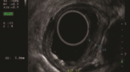
NGHIÊN CỨU ĐẶC ĐIỂM HÌNH ẢNH SIÊU ÂM NỘI SOI Ở BỆNH NHÂN UNG THƯ THỰC QUẢN
13/10/2023 12:07:09 | 0 binh luận
SUMMARY Purpose: The aim of study was decribed the imaging characteristics of endoscopic ultrasound in esophageal cancer. Methods and materials: A cross-sectional descriptive study on 40 patients with esophageal cancer were assigned to endoscopic ultrasound. Results: All study subjects were male with mean age 58.33 ± 7.24. 97.5% of SCC; 2.5% were high-grade dysplasia, none of the patients had adenocarcinoma. Moderately differentiated dominate 55%, 25% highly differentiated and 20% poorly differentiated. .Location of lesions on endoscopy is common in the middle and lower thirds of the esophagus. 85% of esophageal cancer lesions are hypoechoic, 7.5% hyperechoic, and 7.5% mixed. Of which, 70% are heterogeneous, the remaining are homogenous. The lesion size is less than half the circumference accounting for 62.5% and larger than half the circumference accounting for 32.5%. TNM classification on endoscopic ultrasound according to AJCC 8th stage T: Tis (7.5%), T1a (45%), T1b (30), T2 (10%), T3(7.5%). Stage N: N0 (70%), N1 (27.5%), N2 (2.5%). TNM stage: Stage 0 (5%), IA (15%), IB (47.5%), IIB (25%), IIIA (5%), IIIB (2.5%). Conclusion: Endoscopic ultrasound plays an important role in diagnosis and staging in patients with esophageal cancer Key words: endoscopic ultrasound, esophageal cancer, squamous cell carcinoma (SCC), adenocarcinoma.
AN TOÀN VÀ HIỆU QUẢ CỦA ĐỐT NHÂN GIÁP LỚN LÀNH TÍNH BẰNG VI SÓNG (MWA)
12/10/2023 15:30:03 | 0 binh luận
SUMMARY Most thyroid nodules are benign and do not require treatment, but large (≥3 cm) benign thyroid nodules usually cause clinical symptoms or cosmetic concerns and therefore require treatment. Traditionally the treatment for benign thyroid nodules consists of surgery and levothyroxine medication, however, both have many drawbacks. Minimally invasive surgical alternatives are becoming more attractive for the treatment of thyroid nodules. Beside Radiofrequency ablation (RFA), Microwave ablation (MWA) is a method of thermal ablation to induce thyroid tissue necrosis and has been applied to various benign and malignant tumors with good results. Objectives: We determine the safety and efficacy of the Microwave Ablation procedure to treat large benign thyroid nodules. Materials and Methods: We report a series of 40 large benign thyroid nodules patients who underwent Microwave Ablation for treatment at Thoracic Surgery Department Cho Ray hospital from Apr-2018 to Sep-2019. Results: There were 31 females and 9 males, with a median age of 46 years. The medians with the largest diameter and volume of the nodules were 40 mm and 22 ml. Four (10%) minor complications were observed. The mean volume reduction ratio (VRR) was 75.1, 85.2, and 96.4% after 3, 6, and 12 months. The mean symptom and cosmetic scores dropped from 8.0 and 2.8 (before treatment) to 2.8 and 1.3 (at 12 months), respectively. Thirteen nodules (31%) require two MWA sessions. Conclusions: MWA is safe, effective, and can be a good option to treat large benign thyroid nodules. More studies with large datasets and long follow-ups are needed to improve its safety and efficacy. Keywords: Large benign thyroid nodules; Microwave ablation (MWA); Volume reduction ratio; Symptom score; Cosmetic score.
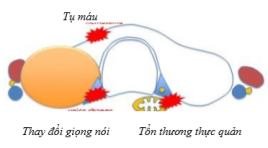
ĐÁNH GIÁ KẾT QUẢ NÚT TẮC ỐNG NGỰC ĐIỀU TRỊ BIẾN CHỨNG RÒ DƯỠNG CHẤP SAU MỔ UNG THƯ TUYẾN GIÁP
11/10/2023 16:24:55 | 0 binh luận
SUMMARY Background: chylous leakage after operation of head and neck is rare but well-known complication. Almost patients with this complication can be treated conservatively but in patients with high flow leakage, the following treatments will be very complicated. Purpose : to report the results of percutaneous thoracic duct embolization (TDE) treatment for chylous leakage of the neck in patients post thyroidectomy and cervical lymph node dissection due to thyroid cancer. Methods : 15 consecutive patients with high flow chylous leak post thyroidectomy were sent to our hospital after failed conservative treatment. All patient were undergone intra nodal lymphagiography then thoracic duct embolization. Results: Fifteen patients with cervical chylorrhea through drainage more than 300 ml/day during average 2 weeks (1 to 5 weeks) were included in this study. TDE was archived in 15/15 patients in which 14/15 TD were embolized ategrade and 1/15 TD was embolized retrograde. One patient had recurrent chylous leakage after 1 week due to recanalization of TD. She was then undergone TD sclerosis injection under CT scanner guidance. All patients had clinical successful with no chylorrhea after intervention. No major complication was noted. All patients were discharged in following weeks after intervention. Conclusion: TDE is minimal invasive and effective treatment for cervical chylous leakage post thyroidectomy. Keywords: chylous leak, chylorrhea, thoracic duct embolization
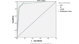
SO SÁNH GIÁ TRỊ CÁC PHÂN LOẠI EU-TIRADS, K-TIRADS VÀ ACR-TIRADS TRONG CHẨN ĐOÁN TỔN THƯƠNG DẠNG NỐT TUYẾN GIÁP
15/11/2021 17:22:56 | 0 binh luận
SUMMARY Purpose: To compare the ultrasound results of thyroid nodules in the application of EU-TIRADS, K-TIRADS, ACR-TIRADS systems with post-operative histological results. To compare diagnostic values of the three TIRADS systems. Material and methods: This is a cross-sectional study with convenient sampling. We recruited thyroid nodules that were performed pre-operative ultrasound (applied EU-TIRADS, K-TIRADS, ACRTIRADS systems) and removal surgery in Hue University of Medicine and Pharmacy Hospital between September 2019 and July 2020. Results: 138 thyroid nodules of 122 patients were enrolled. The malignancy rate was 22.5%. The majority of the lesions were classified as EU-TIRADS 3 (47.8%), K-TIRADS 3 (47.8%) và ACR-TIRADS 2 (38.4%.). The AUC of the EU-TIRADS, K-TIRADS và ACR-TIRADS systems were 0.957, 0.951 and 0.956, respectively. Among the three systems, EU-TIRADS had the highest sensitivity (Se) and negative predictive value (NPV) (100%) while its specificity (Sp), positive predictive value (PPV) and accuracy (Acc) for malignancy were lowest. K-TIRADS showed the best Sp, PPV and Ac (97.2%; 89.7% and 94.2%, respectively) and the lowest other values. Conclusion: The ability to distinguish between the malignant thyroid nodules and the benign ones of the three TIRADS systems was at a very good level. EU-TIRADS showed the most effective diagnostic performance in Se and NPV, while K-TIRADS yielded the best Sp, PPV and Acc. Key words: thyroid nodules, diagnostic values, EU-TIRADS, K-TIRADS, ACR-TIRADS.
Bạn Đọc Quan tâm
Sự kiện sắp diễn ra

THÔNG BÁO SINH HOẠT KHOA HỌC 19/01/2024: CẬP NHẬT CHẨN ĐOÁN VÀ ĐIỀU TRỊ UNG THƯ TRỰC TRÀNG
19-01-2024 09:47:01 0 bình luận

Thông tin đào tạo
- Những cạm bẫy trong CĐHA vú và vai trò của trí tuệ nhân tạo
- Hội thảo trực tuyến "Cắt lớp vi tính đếm Photon: từ lý thuyết tới thực tiễn lâm sàng”
- CHƯƠNG TRÌNH ĐÀO TẠO LIÊN TỤC VỀ HÌNH ẢNH HỌC THẦN KINH: BÀI 3: U não trong trục
- Danh sách học viên đạt chứng chỉ CME khóa học "Cập nhật RSNA 2021: Công nghệ mới trong Kỷ nguyên mới"
- Danh sách học viên đạt chứng chỉ CME khóa học "Đánh giá chức năng thất phải trên siêu âm đánh dấu mô cơ tim"


Đơn vị hợp tác

