


NGHIÊN CỨU ĐẶC ĐIỂM HÌNH ẢNH VÀ VAI TRÒ CỦA SIÊU ÂM TRONG CHẨN ĐOÁN U TUYẾN CẬN GIÁP
13/10/2023 16:21:43 | 0 binh luận
SUMMARY Results: There were 33 patients with primary hyperparathyroidism who had available preoperative ultrasounds of the parathyroid glands. At surgery, 42 parathyroid adenomas were removed in the 33 patients. Ultrasound correctly revealed 38 adenomas in 42 parathyroid adenomas (90.47%). The most common parathyroid adenomas were located on the posterior of the thyroid lobes (97.4%). All of parathyroids adenoma had well-defined margins (100%) and most showed hypoechogenicity (71.1%), and peripheral vascularity was the most common pattern (92.1%). Conclusion: The most common ultrasonographic findings of parathyroid adenomas was well-defined hypoechoic lesion with peripheral vascularity. Ultrasound plays role in pre-operative localization of the parathyroid tumors and supporting clinical diagnosis. Keywords: ultrasound, primary hyperparathyroidism, parathyroid adenoma.
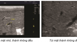
Trường hợp lâm sàng: U cơ tuyến túi mật
20/11/2019 16:01:17 | 0 binh luận
Bệnh nhân nữ 37 tuổi, đi kiểm tra sức khỏe. Tiền căn: không có tiền căn bệnh lý Các xét nghiệm SGOT, SGPT, bilirubin, CTM, chức năng thận trong giới hạn bình thường
Nhân một vài trường hợp điều trị viêm gân vôi hóa bằng chọc hút vôi dưới hướng dẫn siêu âm
02/06/2020 10:39:12 | 0 binh luận
Report of several cases of treatment of calcific tendonitis by aspiration under ultrasound guidance SUMMARY Calcific tendonitis is a common disease caused by the deposition of canxi hydroxyapatite crystals in the tendons. The disease can occur in all tendons in the body and also in the ligaments, but the most common sites are the tendons of rotator cuffs , tendons around great trochanter, tendons around elbow joints, wrists... Normally, there is no pain. However, as calcification resorption occurs because the body releases enzymes that resolve calcification, patients develop severe and persistent pain. In terms of diagnosis, clinical symptoms are quite difficult to distinguish from other causes of musculoskeletal pain, but diagnostic imaging is easy with methods such as radiography, ultrasound, computer tomography, resonance imaging... On the treatment side, medical therapy is the first-line treatment with nonsteroidal anti-inflammatory painkillers. However, in fact, we found that there are many cases of persistant pain that resistant to NSAIDS drugs, because the calcific deposits are quite large (size up to 1-2 cm) so that the calcification resorption process persists for a long time. Percutaneous aspiration of calcification under ultrasound guidance is a minimally invasive treatment that reduces the progression of the disease due to nearly completely calcific aspiration. This technique is quite easy to implement and can be widely disseminated. We have also performed this technique for some patients diagnosed with calcific tendonitis at Hanoi Medical University Hospital and had achieve good clinical efficacy.
Siêu âm phát hiện giãn thực quản trong co thắt tâm vị báo cáo 3 trường hợp
02/06/2020 09:55:31 | 1 binh luận
Ultrasound imaging the dilate esophagus in achalasia reported 3 cases SUMMARY The esophagus can be divided into four sections: the neck, the thoracic, the diaphragm, and the abdominal segment. During the ultrasound examination of patients at the Department of Functional Ultrasound, we found that the sonographer could observe the images of the diaphragmatic esophagus with normal and dilated images. weirdo. Through the study of 3 cases of patients with esophageal dilatation in pathological spasms, we found that the image of esophageal dilatation on the ultrasound is: size> 20 mm, the last segment has a image that can be called "bird beak”, patients who drink water observe that the fluid in their esophagus swirling can be called a “washing machine”sign.
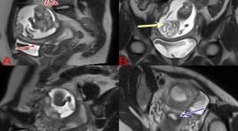
Thai 18 tuần ở sừng chột của tử cung 1 sừng
15/06/2020 11:19:35 | 0 binh luận
A 18 WEEK OF GESTATIONAL AGE IN A NONCOMMUNICATING RUDIMENTARY HORN OF A UNICORNUATE UTERUS
Nghiên cứu giá trị siêu âm trong chẩn đoán teo mật bẩm sinh ở trẻ < 4 tháng
26/03/2020 22:51:55 | 0 binh luận
Value of Ultrasound in diagnosis of biliary atresia in infants younger than 4 months old SUMMARY Purpose: To evaluate prospectively the value of ultrasonography (US) in the diagnosis of biliary atresia (BA), with surgery as the reference standard. Material and Methods : 98 fasting infants (< 4 months old) with jaundice, acholic stools and conjugated hyperbilirubinemia underwent detailed US studies performed by an experienced pediatric radiologist with a 5MHz curvilinear transducer and a 7.5MHz linear-array transducer. The following features were prospectively recorded: gallbladder morphology, size and contraction, triangular cord sign. The radiologist was blinded to results of other investigations. Sensitivity, specificity, and positive and negative predictive values were calculated for each US variable. BA and non-BA groups were compared by means of the Fisher exact test for categorical variables and an unpaired t test for continuous variables. Result : Forty infants had surgically confirmed BA, and 58 had other documented causes of neonatal jaundice; the mean ages at US assessment were 57 and 58 days, respectively (P>0,5). Seven US features showed a significant difference between BA and non-BA groups (P< 0,01, Fisher exact test). The features with the greatest individual sensitivity and specificity, respectively, in the diagnosis of BA were triangular cord sign (87,5% and 94,8%), abnormal gallbladder wall (87,5% and 89,6%) and no contraction (90,6% and 89,6%).The gallbladder was significantly smaller in infants with BA than in those without BA (15,4mm vs 22,5 mm in length, P <0,01). Conclusion : US is valuable in diagnosis of biliary atresia if patients fast enough. Multiple US features should be used to increase the accuracy of the diagnosis Abbreviation : US: Ultrasound BA: Biliary Atresia TC: Triangular Cord Key word: Billiary atresia, Triangle Cord
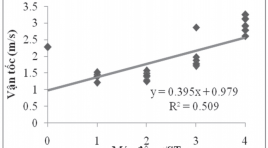
Gía trị vận tốc sóng biến dạng trong mô gan xơ qua kỉ thuật xung áp lực nén siêu âm
03/04/2020 09:33:44 | 0 binh luận
The value of shear wave average velocity in healthy’s liver by using acoustic radiation force impulse imaging SUMMARY: Objectives: The purposes of this study were to measure the reference value of average velocity of shear wave in healthy’s liver by using acoustic radiation force impulse imaging. Methods: Two hundred and of healthy volunteers with normal liver function test values were selected for the study. Shear wave velocity measurements, expressed in meters per second, were taken in liver segment 7 or 8 at depth from 3 to 4cm below the body surface portion of. Among these volunteers, we chose fifty ones to whom two observers with different levels of experience performed the measurements independently and blindly. Results: Total the measurements are taken 2.532 times. The value of average velocity of shear wave in the health’s liver is 1.05 ± 0.12m/s. There is no statistically significant difference in shear wave velocity between two genders. In term of interobserver results, there is no statistically significant difference in shear wave velocity obtained by two observers with different levels of experiences (P < .005). Conclusions: The results of this study show that shear wave velocity measurement in health’s liver by using the acoustic radiation force impulse technique is about 1.05 ± 0.12m/s and this technique is reproducible and independent to user.
Bạn Đọc Quan tâm
- Ca lâm sàng: điều trị ung thư tuyến giáp bằng I-131
- Những tiến bộ trong chẩn đoán hình ảnh bệnh tim – mạch
- Ứng dụng CNTT trong chẩn đoán hình ảnh giúp tăng chất lượng y tế
- CA LÂM SÀNG: UNG THƯ BIỂU MÔ NỘI ỐNG TUYẾN VÚ
- KẾT QUẢ ỨNG DỤNG XẠ HÌNH THẬN ĐỘNG TẠI BỆNH VIỆN NHI ĐỒNG THÀNH PHỐ
- NGHIÊN CỨU BƯỚC ĐẦU ĐÁNH GIÁ VAI TRÒ CỦA TRÍ TUỆ NHÂN TẠO TRONG PHÂN TÍCH KẾT QUẢ X QUANG NGỰC THẲNG TẠI BỆNH VIỆN CHỢ RẪY
- NGHIÊN CỨU ĐẶC ĐIỂM HÌNH ẢNH VÀ VAI TRÒ CỦA SIÊU ÂM TRONG CHẨN ĐOÁN U TUYẾN CẬN GIÁP
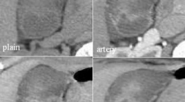
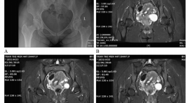
- ĐÁNH GIÁ KẾT QUẢ BƯỚC ĐẦU ĐIỀU TRỊ TĂNG SẢN LÀNH TÍNH TUYẾN TIỀN LIỆT CÓ BÍ TIỂU CẤP BẰNG PHƯƠNG PHÁP CAN THIỆP NÚT ĐỘNG MẠCH TUYẾN TIỀN LIỆT
- VỠ TÚI GIẢ PHÌNH KHỔNG LỒ TÂM THẤT TRÁI: BÁO CÁO 1 TRƯỜNG HỢP HIẾM GẶP TẠI BỆNH VIỆN BẠCH MAI
- VAI TRÒ CỦA SINH THIẾT BẰNG KIM LÕI DƯỚI HƯỚNG DẪN SIÊU ÂM TRONG ĐÁNH GIÁ HẠCH CỔ BẤT THƯỜNG
Sự kiện sắp diễn ra

THÔNG BÁO SINH HOẠT KHOA HỌC 19/01/2024: CẬP NHẬT CHẨN ĐOÁN VÀ ĐIỀU TRỊ UNG THƯ TRỰC TRÀNG
19-01-2024 09:47:01 0 bình luận

Thông tin đào tạo
- Những cạm bẫy trong CĐHA vú và vai trò của trí tuệ nhân tạo
- Hội thảo trực tuyến "Cắt lớp vi tính đếm Photon: từ lý thuyết tới thực tiễn lâm sàng”
- CHƯƠNG TRÌNH ĐÀO TẠO LIÊN TỤC VỀ HÌNH ẢNH HỌC THẦN KINH: BÀI 3: U não trong trục
- Danh sách học viên đạt chứng chỉ CME khóa học "Cập nhật RSNA 2021: Công nghệ mới trong Kỷ nguyên mới"
- Danh sách học viên đạt chứng chỉ CME khóa học "Đánh giá chức năng thất phải trên siêu âm đánh dấu mô cơ tim"


Đơn vị hợp tác

