


VAI TRÒ CỦA PET/CT TRONG UNG THƯ VÚ
23/01/2024 10:42:11 | 0 binh luận
TÓM TẮT Giới thiệu: Ung thư vú là ung thư hàng đầu ở nữ giới. Chẩn đoán chính xác giai đoạn giúp điều trị bệnh nhân một cách hiệu quả và có thể kéo dài thời gian sống cho bệnh nhân. PET/CT là một phương tiện hiệu quả trong chẩn đoán và phân lập giai đoạn ung thư vú. Mục tiêu: Nghiên cứu này xác định vai trò thay đổi giai đoạn ung thư vú sau khi chụp PET/CT, đồng thời đánh giá mức độ hấp thu FDG theo chỉ số SULmax của các tổn thương. Đối tượng và phương pháp: Đối tượng nghiên cứu là các bệnh nhân ung thư vú chụp PET/CT trong năm 2020. Những bệnh nhân đươc loại khỏi nghiên cứu gồm những bệnh nhân rơi vào các tình huống sau: ung thư vú 2 bên, có các bệnh ung thư khác, thiếu các thông tin về giai đoạn bệnh, đường huyết khi tiêm phóng xạ lớn hơn 200 mg/dL. Liều FDG dùng cho các bệnh nhân là 0,1mCi/ kg cân nặng. Chụp PET/CT scan từ đỉnh đầu đến 1/3 trên đùi. Dữ liệu được ghi nhận trước và sau khi chụp. Xử lý dữ liệu với MS Excel và SPSS 25.0. Phương pháp nghiên cứu : hồi cứu, mô tả hàng loạt ca Kết quả: Sau khi sàng lọc, có 80 bệnh nhân được chọn vào nghiên cứu. Trong đó, 47,5% (38 ca) là các trường hợp ung thư vú trái và 52,5% (42 ca) là các ung thư vú phải. Mục đích chụp PET/CT có 33,8% (27 ca) chụp để chẩn đoán giai đoạn và 66.3 % (53 ca) đánh giá tái phát, di căn. Sự thay đổi giai đoạn sau chụp PET/CT được ghi nhận là 47,5% ( 38 ca) với 45% (36 ca) tăng giai đoạn và 2,5% (2 ca) giảm giai đoạn. Khi đánh giá mức độ hấp thu FDG theo SULmax ghi nhận các bướu nguyên phát cho hấp thu cao nhất. Trong các phân nhóm sinh học, phân nhóm tam âm có mức hấp thu trung bình cao nhất. Về các tổn thương di căn phát hiện được thì di căn phổi – màng phổi chiếm tỉ lệ cao nhất với 38,25% (13 ca). Ngoài ra, còn phát hiện 26,27 % (09 ca) các trường hợp di căn từ hai vị trí trở lên. Kết luận: PET/CT đã giúp cho gần một nửa số bệnh nhân được phân loại giai đoạn chính xác và nhận được điều trị đúng mức. Đồng thời các bệnh nhân còn lại cũng được an tâm điều trị theo chiến lược hiện tại. PET/CT còn giúp đánh giá mức độ chuyển hoá glucose của các tổn thương ác tính hoặc phát hiện sớm các tổn thương tái phát, di căn khi chưa có thay đổi về hình thái. Từ khoá: PET/CT, ung thư vú, đánh giá tái phát di căn, BV Ung Bướu
VAI TRÒ CỦA FDG PET/CT TRONG CHẨN ĐOÁN VÀ TIÊN LƯỢNG U PHYLLODES TUYẾN VÚ DI CĂN VÀ HỒI CỨU Y VĂN
18/10/2023 12:01:27 | 0 binh luận
SUMMARY Metastasis of malignant phyllodes tumors is extremely rare that there are only few cases reported in literature. With the aim of providing data and diagnostic guidance, discussing treatments, we present the role of PET/CT, other radiologic modalites. We report a clinical case a 43 years-old female patient who was diagnosed with grade II phyllodes tumor of the breast and treated by surgery and radiotherapy adjuvant (2017). Following up on radiology test after 6 years (02/2023), the patient has diagnosed with bilateral lungs metastasis and left upper lobectomy was done. PET/CT scan in Choray hospital after the surgery showed multiple hypermetabolic foci in lungs, thoracic wall, sternum, pancreas, stomach, bones which suggested distant metastases from the prior tumor. Treatments for distant recurrence phyllodes tumors have not been effective and patients often have poor prognosis. However, some new target therapies may help increase life expectancy, improve quality of life.
ĐÁNH GIÁ KẾT QUẢ SỚM ĐIỀU TRỊ NANG TUYẾN VÚ BẰNG VI SÓNG TẠI ĐƠN VỊ TUYẾN VÚ BỆNH VIỆN CHỢ RẪY
12/10/2023 12:34:22 | 0 binh luận
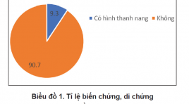
SUMMARY Background- Objective : Breast cyst is common in premenopausal women (61.5 %) in comparison with the menopause population (39.4%) and the most common age is 35 to 50. The treatment of breast cysts using microwave has a lot of advantages and should be a considered method in clinical practice. Our objective is to evaluate the early result of breast cyst treatment with microwave under ultrasound guidance in the breast unit of Cho Ray hospital. Method: We prospective reviewed patients who underwent breast cyst treatment using microwave ablation in Breast unit of Cho Ray hospital Ho Chi Minh city from 08/2018 to 03/2020. Result: 58 patients are reviewed. The average age is 45.3. Simple breast cyst, complicated breast cyst, and complex breast cyst are 80%, 16.93 %, and 3.07 % prospectively. The average diameter of ablated cysts is 23.86 mm. All of the patients have a complete response after treatment, and the decrease in size ratio has reached more than 90 % after 6 months. No complication is recorded in the treatment process. There is one case, with calcification of the cyst wall, that does not respond to microwave ablation. Conclusion: The microwave ablation procedure is a promising method for symptom breast cyst with a high rate of success and low rate of complication. Breast cyst with calcified wall should have a careful assessment before the procedure. Key Word: Breast cyst, Microwave ablation
KẾT QUẢ BAN ĐẦU SINH THIẾT VÚ HÚT CHÂN KHÔNG DƯỚI HƯỚNG DẪN SIÊU ÂM CÓ KẾT HỢP ĐỊNH VỊ KIM CHO TỔN THƯƠNG VI VÔI HÓA
11/10/2023 14:38:52 | 0 binh luận
SUMMARY Purpose: This study examined the usefulness of ultrasound-guided vacuum-assisted breast biopsy for mammographic microcalcification. Methods : case series from June- 2019 to June-2020 at Breast department Cho Ray hospital. The patients with BI-RADS Category 4 Mammographic microcalcification were included. Most microcalcifications were not observed on ultrasound. Sono-guided J-wire localization was first performed for the suspicious microcalcification area, and the location of the J-wire and calcification was determined with mammography in most cases. Sono-guided VABB was performed after removing the J-wire without a stereotactic device. On the other hand, Sono-guided VABB was performed directly without J-wire localization when microcalcification lesions were identified by mass on ultrasonography. In all cases, calcification was confirmed by specimen mammography and the pathology was performed. A follow-up examination was performed to confirm the presence of complications. Results: A total of 20 lesions of 18 patients with BI-RADS Category 4 Mammographic microcalcification were included. Mean age: 49,44 ± 9,49 (35-66). Mean size of lesions 10,83 ± 3,60 mm, (4-15mm). In 20 lesions, 6 lesions (30%) were diagnosed as a malignancy (2 cases of ductal carcinoma in situ, 3 cases of ductal carcinoma invasive, 1 case of atypical ductal hyperplasia). The remaining 14 lesions (70%) were diagnosed as benign (fibroadenoma: 4; fibrocystic exchange 7, fibrocystic desease: 1, typical hyperplasia : 2). There were no significant complications during follow up after VABB. Conclusion: Sono-guided VABB can be used effectively if combined with wire localization for microcalcification lesions. Keywords: Mamography, Calcification, VABB, Image-guides biopsy, J Wire localization
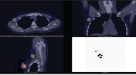
BÁO CÁO 2 TRƯỜNG HỢP SPECT/CT HẠCH GÁC TRONG UNG THƯ VÚ
16/11/2021 11:30:55 | 0 binh luận
SUMMARY Sentinel lymph node is defined as the first lymph node or group of first nodes to which cancer cells are likely to spread from the primary tumor. Cancer cells are often found in sentinel lymph node(s) before metastasizing to other nodes or organs. Therefore, sentinel lymph node biopsy plays an important role in identification and management of metastasis lesions, especially in breast cancer, testicular cancer and melanoma. Sentinel lymph node scintigraphy with SPECT/CT using 99mTechnetiumnanocolloid is a useful imaging technique for nodal determination and localization. With negative histopathological results of sentinel lymph node dissection, axillary lymph node dissection can be avoided for patients thereby reducing the risk of lymphedema and other complications. In this article, two patients in Vinmec Health Care System, Times City with breast cancer who underwent SPECT/CT sentinel lymph node scintigraphy were reported. Keywords: breast cancer, sentinel lymph node, SPECT/CT, 99mTc-nanocolloid
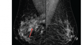
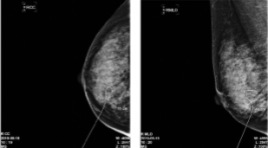
NGHIÊN CỨU GIÁ TRỊ CỦA X-QUANG CẮT LỚP TRONG CHẨN ĐOÁN UNG THƯ VÚ Ở BỆNH NHÂN CÓ VÚ ĐẶC HOẶC BẤT XỨNG KHU TRÚ
16/11/2021 09:56:31 | 0 binh luận
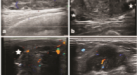
ĐÁNH GIÁ HIỆU QUẢ CỦA CHỌC HÚT ÁP XE VÚ DƯỚI HƯỚNG DẪN SIÊU ÂM Ở PHỤ NỮ CHO CON BÚ
15/11/2021 17:34:43 | 0 binh luận
SUMMARY Objective: To describe the results of ultrasound- guided needle aspiration in the treatment of lactational breast abscesses. Subjects and methods of study: this study was conducted in Bach Mai Hospital, from 6/2020 to 1/2021. Patients with lactating breast abscesses underwent ultrasound- guided aspiration followed by antibiotics therapy. Results: In 34 patients with 46 lactating breast abscesses, most of the abscesses had heterogeneous echogenic, no capsule and size smaller than 5cm. The smallest size of abscesses was 13x24x14mm (equivalent to volume of 2ml), the largest one occupied most of the mammary gland, equivalent to a volume of 540ml. Bacterial culture results showed that 84,8% of cases were methicillin-resistant staphylococcus aureus. The average number of aspirations was 2. The average treatment length was 16 days. Success rate was 91,2% and in these cases, 4 patients (9,3%) had galactocele complication after treatment. 3 cases (8,8%) were converted to the traditional incision and drainage. Conclusion: Ultrasound- guided needle aspiration is an effective and minimally invasive treatment option for lactating breast abscess with a high rate of success and good cosmetic results. Keywords: lactating breast abscess, ultrasound- guided needle aspiration.
GIÁ TRỊ CÁC THÔNG SỐ BÁN ĐỊNH LƯỢNG CỦA CỘNG HƯỞNG TỪ ĐỘNG HỌC TRONG CHẨN ĐOÁN PHÂN BIỆT TỔN THƯƠNG VÚ LÀNH TÍNH VÀ ÁC TÍNH
15/11/2021 17:08:47 | 0 binh luận
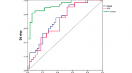
SUMMARY Objective: The aims of this study were to determine the value of the semi-quantitative parameters obtained by dynamic contrast enhancement magnetic resonance imaging (DCE-MRI) in differentiation between benign and malignant breast lesions. Methods: A retrospective study was performed on 63 females (with 72 breast lesions) underwent DCE-MRI before treatment at Cho Ray hospital from Jan 2019 to Feb 2020. The value of semi-quantitative parameters (signal intensity slope (SIslope), maximum slope of increase (MSI), percentage of peak enhancement (Epeak)) were evaluated. The diagnostic value of the time intensity curve according to 5th edition ACR (2013) was compared with the value based on semi-quantitative methods. The results of each DCE-MRI parameter were correlated with histopathology. Results: There were 63 patients with 72 breast lesions including 40 benign lesions and 32 malignant lesions. The area under the ROC curve of SIslope, MSI and Epeak were 0,908; 0,702 and 0,734, respectively. The sensitivity, specificity and accuracy of the time intensity curve according to ACR and semi-quantitative methods were 68,8% and 87,5%; 87,5% and 85%; 79,2% and 86,1%, respectively. Conclusion: Our study reinforces the importance of the semi-quantitative parameters of DCE-MRI in distinguishing between benign and malignant breast lesions. Semi-quantitative analysis of the time intensity curve helps to increase the diagnostic accuracy compared with the methods of ACR. Key words: breast lesions; semi-quantitative parameters; time intensity curve.
Bạn Đọc Quan tâm
- Ca lâm sàng: điều trị ung thư tuyến giáp bằng I-131
- Những tiến bộ trong chẩn đoán hình ảnh bệnh tim – mạch
- Ứng dụng CNTT trong chẩn đoán hình ảnh giúp tăng chất lượng y tế
- CA LÂM SÀNG: UNG THƯ BIỂU MÔ NỘI ỐNG TUYẾN VÚ
- KẾT QUẢ ỨNG DỤNG XẠ HÌNH THẬN ĐỘNG TẠI BỆNH VIỆN NHI ĐỒNG THÀNH PHỐ
- NGHIÊN CỨU BƯỚC ĐẦU ĐÁNH GIÁ VAI TRÒ CỦA TRÍ TUỆ NHÂN TẠO TRONG PHÂN TÍCH KẾT QUẢ X QUANG NGỰC THẲNG TẠI BỆNH VIỆN CHỢ RẪY
- NGHIÊN CỨU ĐẶC ĐIỂM HÌNH ẢNH VÀ VAI TRÒ CỦA SIÊU ÂM TRONG CHẨN ĐOÁN U TUYẾN CẬN GIÁP
- ĐÁNH GIÁ KẾT QUẢ BƯỚC ĐẦU ĐIỀU TRỊ TĂNG SẢN LÀNH TÍNH TUYẾN TIỀN LIỆT CÓ BÍ TIỂU CẤP BẰNG PHƯƠNG PHÁP CAN THIỆP NÚT ĐỘNG MẠCH TUYẾN TIỀN LIỆT
- VỠ TÚI GIẢ PHÌNH KHỔNG LỒ TÂM THẤT TRÁI: BÁO CÁO 1 TRƯỜNG HỢP HIẾM GẶP TẠI BỆNH VIỆN BẠCH MAI
- VAI TRÒ CỦA SINH THIẾT BẰNG KIM LÕI DƯỚI HƯỚNG DẪN SIÊU ÂM TRONG ĐÁNH GIÁ HẠCH CỔ BẤT THƯỜNG
Sự kiện sắp diễn ra

THÔNG BÁO SINH HOẠT KHOA HỌC 19/01/2024: CẬP NHẬT CHẨN ĐOÁN VÀ ĐIỀU TRỊ UNG THƯ TRỰC TRÀNG
19-01-2024 09:47:01 0 bình luận

Thông tin đào tạo
- Những cạm bẫy trong CĐHA vú và vai trò của trí tuệ nhân tạo
- Hội thảo trực tuyến "Cắt lớp vi tính đếm Photon: từ lý thuyết tới thực tiễn lâm sàng”
- CHƯƠNG TRÌNH ĐÀO TẠO LIÊN TỤC VỀ HÌNH ẢNH HỌC THẦN KINH: BÀI 3: U não trong trục
- Danh sách học viên đạt chứng chỉ CME khóa học "Cập nhật RSNA 2021: Công nghệ mới trong Kỷ nguyên mới"
- Danh sách học viên đạt chứng chỉ CME khóa học "Đánh giá chức năng thất phải trên siêu âm đánh dấu mô cơ tim"


Đơn vị hợp tác

