


Cập nhật vai trò của chẩn đoán hình ảnh trong chẩn đoán viêm phổi do SARS-CoV2
25/08/2021 12:57:39 | 0 binh luận
Vai trò của 3D TOF MRA trong đánh giá rò động tnh mạch màng cứng nội sọ
25/08/2021 13:00:13 | 0 binh luận
Vai trò của chụp cắt lớp vi tnh mạch máu trong đánh giá hẹp tắc động mạch nội sọ ở bệnh nhân đột quỵ do thiếu máu não cấp
25/08/2021 13:03:53 | 0 binh luận
Tương quan giữa hình chụp cắt lớp vi tnh, nội soi với mô bệnh học sau mổ trong phân giai đoạn T ung thư thanh quản
25/08/2021 14:03:47 | 0 binh luận
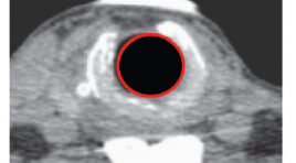
Nghiên cứu vai trò của cắt lớp vi tnh trong chẩn đoán ung thư lưỡi
25/08/2021 14:05:56 | 0 binh luận
Khảo sát đặc điểm hình ảnh trên cắt lớp vi tính 128 dãy có dựng hình ba chiều trong bệnh lý sẹo hẹp khí quản tại Bệnh viện Bạch Mai
06/05/2021 12:36:00 | 0 binh luận
SUMMARY Objective: The study aimed to research causes, imaging features on CT scan and endoscopy, the correlation between CT scan and endoscopy in tracheal stenosis disease. Subjects and methods: Descriptive cross-sectional study, 14 patients was diagnosed tracheal stenosis at Bach Mai hospital from March 2019 to July 2020. Instruments of data collection included medical records, endoscopy and CT scan results. SPSS software was then employed for data analysis. Results: 14 patients in the study, 10 males (71,4%), 4 females (28,6%). Average age was 41.7±13.8. The cause of intubation and tracheostomy was diverse, but the main cause was traumatic brain injuries due to traffic accidents (4 patients). Prominent clinical symptom was difficult inspiration and stridor. Imaging features of tracheal stenosis on CT scan: The mean distance from vocal cord was 29mm. According to Cotton classification, grade III was highest proption ( 71,4%), the remaining was grade II (28,6%), no case was grade I and IV. The mean length of stenosis was 20mm. On endoscopy, the mean distance from vocal cord was 33mm. The correlation between three-dimensional 128-slice CT scanning and endoscopy was concluded: Similar evaluation of the distance from vocal cord to upper end of stenosis, no statistical significance (p>0.05) Key words: tracheal stenosis, intubation and tracheostomy, 128-slice CT scanning, the correlation between CT scan and endoscopy
Bạn Đọc Quan tâm
- Ca lâm sàng: điều trị ung thư tuyến giáp bằng I-131
- Những tiến bộ trong chẩn đoán hình ảnh bệnh tim – mạch
- Ứng dụng CNTT trong chẩn đoán hình ảnh giúp tăng chất lượng y tế
- CA LÂM SÀNG: UNG THƯ BIỂU MÔ NỘI ỐNG TUYẾN VÚ
- KẾT QUẢ ỨNG DỤNG XẠ HÌNH THẬN ĐỘNG TẠI BỆNH VIỆN NHI ĐỒNG THÀNH PHỐ
- NGHIÊN CỨU BƯỚC ĐẦU ĐÁNH GIÁ VAI TRÒ CỦA TRÍ TUỆ NHÂN TẠO TRONG PHÂN TÍCH KẾT QUẢ X QUANG NGỰC THẲNG TẠI BỆNH VIỆN CHỢ RẪY
- NGHIÊN CỨU ĐẶC ĐIỂM HÌNH ẢNH VÀ VAI TRÒ CỦA SIÊU ÂM TRONG CHẨN ĐOÁN U TUYẾN CẬN GIÁP
- ĐÁNH GIÁ KẾT QUẢ BƯỚC ĐẦU ĐIỀU TRỊ TĂNG SẢN LÀNH TÍNH TUYẾN TIỀN LIỆT CÓ BÍ TIỂU CẤP BẰNG PHƯƠNG PHÁP CAN THIỆP NÚT ĐỘNG MẠCH TUYẾN TIỀN LIỆT
- VỠ TÚI GIẢ PHÌNH KHỔNG LỒ TÂM THẤT TRÁI: BÁO CÁO 1 TRƯỜNG HỢP HIẾM GẶP TẠI BỆNH VIỆN BẠCH MAI
- VAI TRÒ CỦA SINH THIẾT BẰNG KIM LÕI DƯỚI HƯỚNG DẪN SIÊU ÂM TRONG ĐÁNH GIÁ HẠCH CỔ BẤT THƯỜNG
Sự kiện sắp diễn ra

THÔNG BÁO SINH HOẠT KHOA HỌC 19/01/2024: CẬP NHẬT CHẨN ĐOÁN VÀ ĐIỀU TRỊ UNG THƯ TRỰC TRÀNG
19-01-2024 09:47:01 0 bình luận

Thông tin đào tạo
- Những cạm bẫy trong CĐHA vú và vai trò của trí tuệ nhân tạo
- Hội thảo trực tuyến "Cắt lớp vi tính đếm Photon: từ lý thuyết tới thực tiễn lâm sàng”
- CHƯƠNG TRÌNH ĐÀO TẠO LIÊN TỤC VỀ HÌNH ẢNH HỌC THẦN KINH: BÀI 3: U não trong trục
- Danh sách học viên đạt chứng chỉ CME khóa học "Cập nhật RSNA 2021: Công nghệ mới trong Kỷ nguyên mới"
- Danh sách học viên đạt chứng chỉ CME khóa học "Đánh giá chức năng thất phải trên siêu âm đánh dấu mô cơ tim"


Đơn vị hợp tác

