


NGHIÊN CỨU ĐẶC ĐIỂM HÌNH ẢNH 18FDG-PET/CT HẠCH CỔ Ở BỆNH NHÂN UNG THƯ VÒM MŨI HỌNG
14/10/2023 11:57:56 | 0 binh luận
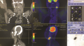
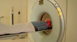
SUMMARY Purpose: Describe imaging characteristics and evaluate the role of 18FDG-PET/CT in the diagnosis of cervical lymph nodes in nasopharyngeal cancer patients. Subjects and methods: Prospective description of 60 patients with nasopharyngeal carcinoma identified by biopsy and histopathology and untreated (in which 55 patients with undifferentiated carcinoma and 5 patients with squamous cell carcinoma), were taken with 18FDG PET/CT and compared with ultrasound and done FNA lymph nodes respectively, from June 2018 to July 2019 to K Central Hospital, Tan Trieu campus. All 18FDG-PET/CT films were read and compared with their respective locations on ultrasound and lymph node FNA before treatment. Results: In total 60 patients common age is 52.3±11.1, male/female ≈ 5/1; A total of 379 bilateral cervical lymph nodes on PET/CT, 195 lymph nodes in the left neck are common (51.5%), the most common lymph node location in group II (96.7% of patients and 61% of total lymph nodes). There are 322/379 (85%) lymph nodes with structural loss on ultrasound, there is a correlation between lymph node size and lymph node absorption on 18FDG-PET/CT with r=0.6, cervical lymph node groups 322/379 Structural abnormal lymph nodes with SUVmax = 9.5 ± 4.6 (nearly 4 times higher than 57/379 normal lymph nodes with SUVmax = 2.6 ± 2.5). In a total of 379 lymph nodes over 60 patients, there was a close agreement between the lymph nodes with loss of umbilical fat structure on ultrasound with SUVmax>2.5 threshold of 92.9% (kappa=0.67) and better at the threshold. SUVmax>3.5 was 92.9% (Cohen's kappa=0.75) on PET/CT. In addition, cytology was positive in 60/89 lymph nodes undergoing FNA, which was in close concordance with ultrasonography of loss of hilar fat structure and cytological diagnosis of 83.4% (Cohen's kappa = 0.62). ). The cytological diagnosis was mild with increased uptake with SUVmax>2.5 threshold of 75.3% (Cohen's kappa = 0.3), but there was a strong correlation when the lymph node imaging was increased. Absorption with SUVmax >2.5 on PET/CT and loss of umbilical cord fat on ultrasound with cytological diagnosis was 87.6% (Cohen's kappa = 0.69). In particular, out of a total of 340/379 lymph nodes with threshold SUVmax>2.5 on PET/CT are considered as metastatic nodes, changing the stage of 25/60 patients (41.67%); in which 17 patients increased and 8 patients decreased stage N compared with staging by other imaging methods before having PET/CT. Conclusion: 18FDG-PET/CT scan is an imaging method with accurate diagnostic value in diagnosing cervical lymph nodes in patients with cervical cancer, useful for treatment planning. Key words: lymph nodes, nasopharyngeal cancer, 18FDG-PET/CT lymph nodes on nasopharngeal cancer.
ĐÁNH GIÁ SỐNG THÊM TRUNG HẠN ĐIỀU TRỊ NÚT MẠCH HÓA CHẤT BỆNH NHÂN UNG THƯ BIỂU MÔ TẾ BÀO GAN TẠI BỆNH VIỆN BẠCH MAI
13/10/2023 15:44:22 | 0 binh luận
SUMMARY Purpose: Evaluation the survival of the patients with hepatocellular carcinoma (HCC) who underwent transcatheter arterial chemoembolization (TACE). Subjects and methods: Retrospective study on transcatheter arterial chemoembolization in patients with hepatocellular carcinoma from July 2017 to July 2020 at Radiology Center - Bach Mai Hospital. Results:. 70 patients including 64 men (92.9%) and 6 women (7.1%) had a confirmed diagnosis of HCC with a mean age of 61.5 ± 11.34 years (from 32 to 83 years). The mean number of treatment sessions were 4.5±0.4 lần (1-16). Progression-free survival after initial TACE and overall survival median was 29.7±3.3 months, 43.3±2.5 months, respectively. Cumulative survival time at 1 year, 2 years, 3 years and 5 years is 91.4%, 77.1%, 63.7% and 29.4%, respectively. Patients treated with cTACE were 67.1% and Deb-TACE were 32.9%. mRECIST after the first TACE: complete response, partial response, stable disease and advanced disease were 30.0%, 55.7%, 4.3%, 10.0%, respectivel. Patients with objective response had a survival of 47.8±2.3 months, and patients with poor response had a survival of 12.2±2.7 months, respectively (p = 0.001). mRECIST response predicted survival among the patients on univariate analysis (HR=15.13, p=0.000). The independent predictors for survival were mRECIST response (p=0.001). Conclusion : Use of TACE in intermediate stage HCC patients gives a significant survival advantage when objective response is achieved as per mRECIST. Key words: TACE, Progression-free survival, overall survival..
ĐÁNH GIÁ PHÂN BỐ DƯỢC CHẤT PHÓNG XẠ 18F-FLUORINE-FLUOROTHYMYDINE TRÊN CHUỘT THÍ NGHIỆM GÂY KHỐI U
13/10/2023 12:49:57 | 0 binh luận
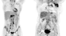
SUMMARY PET (positron emission tomography) techniques and radiopharmaceutical tracers are becoming increasingly common. Since 1991, 3’-deoxy-3’-18Fluorine-Fluorothymidine (18F-FLT) has been studied as a PET tracer for tumor proliferation assessment. The purpose of this study was to assess tracer uptake (%ID/g) in tumor-bearing mice. Materials and methods: Two cell lines (4T1 and LLC) were inoculated into BALB/c mice. The radiotracer was then injected, and tumor resection and organ samples were performed at 20, 40, 60, 90 and 120 minutes later. A gamma-ray spectrometer was used to calculate uptake values. Results: The data show that the tumor’s %ID/g were 3.68 ± 0.29 and 5.30 ± 0.44 at 60-minute post-injection for the 4T1 and LLC tumors, respectively. In other organs, the high uptake was observed in the kidneys, spleen, and bone marrow, but a low level of tracer was seen in the brain. Keywords: 3’-deoxy-3’-18Fluorine-Fluorothymidine, positron emission tomography, tumor-bearing mice.
ĐÁNH GIÁ BƯỚC ĐẦU GIÁ TRỊ CỦA INTERIM PET/CT SAU 2 CHU KỲ ĐIỀU TRỊ HOÁ CHẤT ABVD CỦA BỆNH NHÂN U LYMPHO HODGKIN
13/10/2023 10:44:46 | 0 binh luận
SUMMARY Purpose: The study aimed to initial evaluate the value of interim PET/ CT after 2 cycles of ABVD chemotherapy in Hodgkin lymphoma patients. Material and Methods: The study was carried out retrospective and prospective descriptive study on 56 patients at Tan Trieu K Hospital from March 2020 to June 2022. Patients were examined clinically, paraclinically, had a CT scan or PET/CT film before treatment, then received ABVD chemotherapy, after 2 cycles of being taken and evaluated Interim PET/CT, and continued treatment based on the results. Result of iPET/CT leads to continue or change of regimen continued treatment or change of regimen and follow-up. Results: In a total of 56 patients, male/female ratio: 1/1.6, mean age 30±12.6 (youngest age 7 years old, oldest age 67 years old). 35/56 patients (62.5%) had CT scan and 21/56 patients (37.5%) had background PET/CT scan before treatment. 30.4% had 2 lymph node sites and 28.6 patients had 3 lymph node sites on the body, lymph nodes above the diaphragm 67.9% and lymph nodes both above and below the diaphragm 12,5%. 49 patients had lymph node lesions, short axis size of lymph nodes maximum 27.10±8.3 mm. Lesion to the mediastinum in 15/58 patients (25.8%), spleen in 5 patients. Patients with early stage I-II accounted for 82.1%, advanced stage III-IV accounted for 17.9%. After 2 cycles of ABVD chemotherapy, the response rate on PET/CT was iPET/CT2 (-) 43/56 patients (76.8%, of which Deuville score 1 point 93%) and iPET/CT2 (+) in 13/56 patients (23.3%). The prognostic index of IPS low-risk group (0-2) complete metabolic response was 86.4%. In the high-risk group IPS (4-7) partial metabolic response and complete metabolic response was 54.5% according to Lugano classification. Conclusion: Interim PET/CT has an important role in providing prognostic information in the treatment of ABVD chemotherapy in Hodgkin Lymphoma patients, reducing the toxicity of chemotherapy and determining the next treatment modalities. Keywords: lymphoma Hodgkin, interim PET/CT.
NGHIÊN CỨU ĐẶC ĐIỂM HÌNH ẢNH VÀ VAI TRÒ CỦA 18FDG-PET/CT TRONG CHẨN ĐOÁN GIAI ĐOẠN CỦA BỆNH NHÂN UNG THƯ PHỔI KHÔNG TẾ BÀO NHỎ
12/10/2023 15:07:18 | 0 binh luận
SUMMARY Objectives : Study on 18FDG-PET/CT imaging characteristics of lesions in non-small cell lung cancer before treatment and the role of 18FDG-PET/CT in the staging of patients with non-small cell lung cancer. Methods: Retrospective study, descriptive analysis on 43 non-small cell lung cancer patients who were newly detected taken 18FDG-PET/CT at Military Hospital 103 from February 2017 to February 2022, including 32 patients who were indicated for surgery after 18FDG-PET/CT scan without any specific treatment. Results: The average size of lung tumor was 40.98±21.53mm, there was a correlation between SUVmax index and primary tumor size. The SUVmax value increased with stage T. The mean SUVmax value of the lymph node group >10mm was 10.59±6.12 higher than that of the node group <10mm was 5.56±2.47. After PET/CT scan, the N0 stage accounted for 32.6%, the N1 stage accounted for 23.3%, the N2 stage accounted for 23.3%, and the N3 stage accounted for 20.9%. The mean SUVmax value of the tumor in the M1 group of patients was 15.96±4.29, which was higher than that of the M0 group of patients was 14.57±8.26.18FDG-PET/ CT detected 100% of primary tumor lesions in the lung, 18FDG-PET/CT accurately diagnosed stage T at 71.88%, higher than CT with 68.75%, accurately diagnosed stage T at 68.75%. N segment at 81,25%, higher than CT with 75%. Sensitivity, specificity, positive predictive value, negative predictive value of 18FDG-PET/CT in the diagnosis of stage N are: 100%; 68,42%; 68,42% and 100%. Keywords: Non-small cell lung cancer, 18FDG-PET/CT, SUVmax.
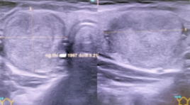
KẾT QUẢ ĐIỀU TRỊ ĐỐT U TUYẾN GIÁP LÀNH TÍNH BẰNG VI SÓNG TẠI VIỆN Y HỌC PHÓNG XẠ VÀ U BƯỚU QUÂN ĐỘI
12/10/2023 12:29:19 | 0 binh luận
SUMMARY Objective: To evaluate the early results of Microwave ablation in the treatment of benign thyroid tumors at the Vietnamese Military Institute Of Medical Radiology And Oncology from 10/2020 to 3/2022. Subjects and Methods : Prospective, descriptive studies of 32 patients with benign thyroid tumors treat by microwave ablation at the Vietnamese Military Institute Of Medical Radiology And Oncology from 10/2020 to 3/2022. Results: Mean of age: 40.91 ± 17.00, female (90,63%), male (9,37%). The solitary tumor occults 62.5%, the thyroid tumors in both glands: are 37.49%, and the average tumor size: is 24.27 ± 7.65 mm. The tumors classified into TIRADS 3 by ultrasound was 96,87%. The mean operating time was 27 ± 5 minutes, all patients were coming home after treatment. Post-operative infection: 3,13%, minor skin burn: 3.13%. The disease symptoms were lost after 3 months of therapy. Conclusions: Using microwave ablation in the treatment of benign thyroid tumors was quite safe, and achieved high cosmetic outcomes. Keyword: Thyroid nodule, Microwave ablation

BƯỚC ĐẦU NGHIÊN CỨU ĐẶC ĐIỂM HÌNH ẢNH VÀ VAI TRÒ CỦA 18FDG-PET/CT TRONG CHẨN ĐOÁN GIAI ĐOẠN UNG THƯ ĐẠI TRỰC TRÀNG TRƯỚC ĐIỀU TRỊ
11/10/2023 16:44:42 | 0 binh luận
SUMMARY Objectives: To evaluate the imaging characteristics and the role of 18FDG-PET/CT in pre-treatment colorectal cancer staging. Subjects and methods : Study on 39 colorectal cancer patients without specific treatment who had taken 18FDG-PET/CT for staging diagnosis, at the Department of Nuclear Medicine, Imaging Center of Military Hospital 103, from February 2017 to November 2020. Results: The average age was 62.77 ± 14.07. The ability to detect primary tumor lesions in the colorectal of 18FDG-PET/CT was 97.44%. The majority of patients had rectal tumors, the tumor size was mainly in the range of 5-10 cm and the T3 stage accounted for the most proportion with 60.5%. There was 46.2% of patients had regional lymph node metastasis, in which the group of lymph nodes over 10 mm and the N2 group accounted for a lower proportion, but the mean SUVmax was statistically significantly higher than others group. Distant metastasis was occured mainly in liver, lung-pleural and bone. There were 59.0% patients in stage III and IV. Diagnosis by 18FDG-PET/CT was accurately at 80.77% for T stage and 66.67% for N stage. The sensitivity and specificity, positive predictive value and negative predictive value of 18FDG-PET/CT in the diagnosis of regional lymph node metastasis were 100% and 60%; 55.55% and 100%. Keywords : Colorectal cancer, 18FDG-PET/CT, SUVmax.
Ứng dụng kỹ thuật PET/CT mô phỏng lập kế hoạch xạ trị ung thư
21/11/2019 08:52:05 | 0 binh luận
Sử dụng PET/CT mô phỏng lập kế hoạch xạ trị gia tốc, đặc biệt IMRT có hiệu quả cao, chính xác, không bỏ sót tổn thương, giảm thiểu tác dụng phụ cơ quan lành, nâng cao chất lượng cuộc sống.
Bạn Đọc Quan tâm
Sự kiện sắp diễn ra

THÔNG BÁO SINH HOẠT KHOA HỌC 19/01/2024: CẬP NHẬT CHẨN ĐOÁN VÀ ĐIỀU TRỊ UNG THƯ TRỰC TRÀNG
19-01-2024 09:47:01 0 bình luận

Thông tin đào tạo
- Những cạm bẫy trong CĐHA vú và vai trò của trí tuệ nhân tạo
- Hội thảo trực tuyến "Cắt lớp vi tính đếm Photon: từ lý thuyết tới thực tiễn lâm sàng”
- CHƯƠNG TRÌNH ĐÀO TẠO LIÊN TỤC VỀ HÌNH ẢNH HỌC THẦN KINH: BÀI 3: U não trong trục
- Danh sách học viên đạt chứng chỉ CME khóa học "Cập nhật RSNA 2021: Công nghệ mới trong Kỷ nguyên mới"
- Danh sách học viên đạt chứng chỉ CME khóa học "Đánh giá chức năng thất phải trên siêu âm đánh dấu mô cơ tim"


Đơn vị hợp tác

