


KẾT QUẢ ĐIỀU TRỊ CAN THIỆP NỘI MẠCH GIẢ PHÌNH ĐỘNG MẠCH TẠNG BỤNG KHÔNG DO CHẤN THƯƠNG
13/10/2023 15:18:32 | 0 binh luận
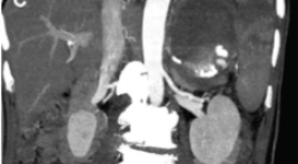
SUMMARY Objective: to apply and estimate the efficacy of embolization in non traumatic visceral arterial pseudoaneurysm. Subject and method: Twenty-seven patients underwent angiography and embolization at Bachmai hospital from 7/2020 to 7/2022. Result: In 27 patients (20 males & 07 females), 09 patients show extravasation in DSA. The splenic artery and the superior mesenteric artery are the arteries most commonly with visceral arterial pseudoaneurysms. Of the 27 patients studied, 24 had technical success in the first intervention, one required secondary intervention because of rebleeding and it was successfully treated by reintervention, one required ileectomy after embolisation due to ongoing hemorrhage, one patient died from uncontrolled blood transfusion shock. Conclusion: In hemodynamically stable and controlled patients, selective and super selective embolization is a safe and effective method for managing non traumatic visceral arterial pseudoaneurysm. Keywords: Visceral arterial pseudoaneurysm (VAPA), embolization, extravasation.
GIÁ TRỊ CỦA CỘNG HƯỞNG TỪ KHUẾCH TÁN TRONG PHÂN BIỆT U MÀNG NÃO ĐIỂN HÌNH VÀ KHÔNG ĐIỂN HÌNH
13/10/2023 12:54:47 | 0 binh luận
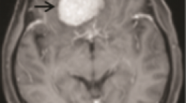
SUMMARY Objective: To determine the value of Diffusion-weighted imaging in distinguishing typical and atypical meningiomas. Subjects and methods: The study was performed at Choray Hospital from January 2021 to August 2022, with 66 typical and atypical meningiomas patients. The descriptive retrospective cross-sectional study was performed. Compare the tumor's signal intensity and mean ADC value with the histopathological results to distinguish typical and atypical meningiomas. Results: In 66 patients, there were 13(19.7%) male patients and 53(80.3%) female patients. The mean age of patients was 60.53 years old. The number of typical and atypical meningiomas cases was 43 (65.2%) and 23 (34.8%). There was no significant signal intensity difference between typical and atypical meningiomas (p=0.56). The mean ADC value for typical meningiomas is 0.843x10⁻³mm2/s and atypical is 0.737x103mm2/s, the difference is statistically significant with p=0.003. On the ROC curve, with an ADC cutoff of 0.780x10⁻³mm2/s, DWI can distinguish typical and atypical meningioma with sensitivity, specificity, PPV, NPV, and accuracy is 83.8%, 51.7%, 67.4%, 71.4%, 66.6% respectively. Conclusion: The mean ADC value of typical meningiomas is higher than that of atypical meningiomas. DWI is valuable in the differential diagnosis of typical and atypical meningiomas. Keywords: meningiomas, typical, atypical, diffusion-weighted imaging, ADC.
NGHIÊN CỨU ĐẶC ĐIỂM HÌNH ẢNH CỘNG HƯỞNG TỪ SỌ NÃO Ở TRẺ SƠ SINH ĐỦ THÁNG CÓ BỆNH LÝ NÃO DO THIẾU MÁU CỤC BỘ/THIẾU OXY
13/10/2023 12:40:53 | 0 binh luận
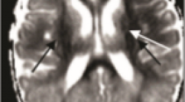
SUMMARY Objectives: to describe the intracranial injuries on brain MRI of term newborns with hypoxic ischemic encephalopathy (HIE) Methods: A prospective and retrospective study based on medical records and brain MRIimages of term newborns with HIE. Brain MRI was performed within 2 weeks after birthon T1, T2, and DWI, SWI sequences Results: There was 94 eligible cases included in the study (male/ female:61/33). The mean gestational age was 39 weeks; mean age of brain MRI was 8 days, mean birth weight was 3100gr. The infants were classified by Sarnat criteria as mild/moderate/severeHIE by ratio: 22.3%/64.9%/12.8%. The infants were mainly given birth by vaginaldelivery 51%, which was followed by cesarean section 45.7%. The sentinel events weredefined mostly as prolonged labor causing fetal distress and placental abruption,umbilical cord accidents. Therapeutic hypothermia was performed in 80,9% cases.Intracranial injury was present in 82 (87.2%) infants. The brain lesions on MRIdiversified. The most common brain injuries were in deep gray nuclei and posterior limbof the internal capsule: thalamus (62.8%), the lenticular nucleus (60.6%), posterior limbof the internal capsule (47.9%), caudal nucleus (26.6%). The following was cortical andsubcortical white matter abnormalities 44.7%. The periventricular and punctate whitematter injuries are less common: 23.4% and 15.9%.The other brain injuries include the corpus callosum (35.1%), the optic radiation(30.8%), hippocampus (12.8%). The infratentorial structures injuries are less common,predominantly in severe HIE cases: brainstem injury 23.1% and cerebellar injury 9.6%.Cortical abnormalities were seen in 42 of 94 (44.7%) predominantly in central sulcus(26.6%), interhemispheric sulcus (21.3%). The cortex injuries in other regions includedinsular region (8.5%), frontal region (27.7%), parietal region (23.4%), occipital region (13.8%), temporal region (8.5%) Intracranial hemorrhage was present in 36/94 (38.3%) infants, including 32 (34%) with subdural hemorrhage, followed by intraventricular hemorrhage (5.3%), intraparenchymal hemorrhage (5.3%) Deep gray nucleus injuries, optic radiation, corpus callosal, hippocampal,brainstem, periventricular and subcortical white matter injuries were significantlyassociated with severity of HIE. The cortical injuries in interhemispheric, insular, frontaland temporal regions were more common in the infants with severe HIE Conclusion: Intracranial injury in the term newborn with HIE diversify, predominantly insupratentorial injuries. The most common brain injuries were in the deep gray nuclei andposterior limb of the internal capsule, followed by cortical and subcortical white matter.The corpus callosum, optic radiation, and hippocampus abnormalities were alsofrequently seen. The brainstem injury is significantly less common and cerebellar injuryis rarely seen. Posterior subdural hemorrhage was the most common intracranialhemorrhage, which was not severe clinically and frequently resolve without treatment Key words: newborn, hypoxic ischemic encephalopathy (HIE), brain MRI Abbreviations: HIE: hypoxic ischemic encephalopathy. MRI: Magnetic Resonance Imaging
ĐÁNH GIÁ HÌNH THÁI TIỂU NHĨ TRÁI TRÊN CẮT LỚP VI TÍNH ĐA DÃY Ở BỆNH NHÂN TRIỆT ĐỐT RUNG NHĨ QUA ỐNG THÔNG
13/10/2023 11:29:50 | 0 binh luận
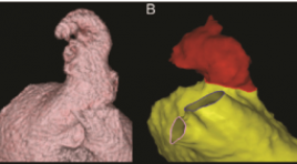
SUMMARY Purpose: to assess left atrial appendage morphology by multidetector computed tomography in atrial fibrillation (AF) patients for catheter ablation Materials and Methods: This retrospective and cross-sectional study included 45 patients diagnosed with AF and treated by catheter ablation and 45 patients without AF undergoing multi-detector computed tomography in E hospital from 1/2020 to 7/2022 Results: The mean age 56.16 ± 11.83, median of left atrium volume in the AF group was 118.13 (96 - 145.56) ml > the control group 72.88 (60.53 - 95.74) ml, (p < 0.001). The most common left atrium morphology in both groups is "cactus" with 46.67% in the AF group and 33.33% in the control group, the middle left atrial appendage orifice was the most common in the AF group (53,33%), while in the control group, the lower left atrial appendage orifice was the most common (44.44%), the length of the left atrial appendage in the AF group was 43.15 ± 8.11 mm, the control group was 44.29 ± 9.76 mm. There is a positive correlation with the mean level between left atrial appendage length and left atrial volume (r = 0.573, p < 0.001). Conclusion: The most common left atrium morphology in both groups is "cactus". There is a positive correlation with the mean level between left atrial appendage length and left atrial volume. Keywords: atrial fibrillation, catheter ablation, left atrial, left atrial appendage, multi-detector computed tomography
BÁO CÁO CA LÂM SÀNG: PHỐI HỢP CHẸN BÓNG ĐỘNG MẠCH CHẬU TRONG VÀ NÚT ĐỘNG MẠCH TỬ CUNG DỰ PHÒNG XUẤT HUYẾT Ở SẢN PHỤ MẮC RAU CÀI RĂNG LƯỢC KẾT HỢP RAU TIỀN ĐẠO
12/10/2023 15:53:35 | 0 binh luận
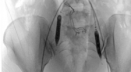
SUMMARY Placenta acrreta spectrum is a condition in which the placenta partially or completely invades and cannot be separated from the uterus muscle. Placenta previa is a condition in which the placenta partially or completely covers the cervix. Both phenomena increase the risk of postpartum haemorrhage, hemostasis disorders, and threaten the life of the mother and the fetus. Combine of the two phenomena increase blood loss in cesarean delivery as well as postpartum period, and is a great challenge required multidisciplinary for successful management. Prophylactic endovascular intervention is a minimally invasive treatment method, which plays an important role in prevent haemorrhage in the management of placenta accreta spectrum. This report describes a case of placenta percreta combine with placenta previa, which received prophylactic endovascular intervention using balloon occlusion of internal illiacs and uterine arteries embolization and underwent a cesarean delivery at 36 weeks. The patient had a successful delivery and preserved the uterus after surgery. Key word : Placenta acrreta spectrum, prophylatic balloon occlusion, uterine artery embolization
SINH THIẾT QUA DA ĐƯỜNG TĨNH MẠCH CẢNH TRONG KẾT HỢP CAN THIỆP ĐIỀU TRỊ HỘI CHỨNG TĨNH MẠCH CHỦ TRÊN: BÁO CÁO TRƯỜNG HỢP LÂM SÀNG
12/10/2023 14:20:08 | 0 binh luận
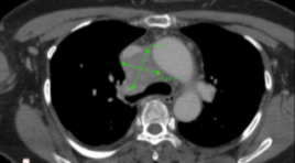
SUMMARY Superior vena cava syndrome often has a mediastinal malignant origin, although it is not commonly found in clinical practice. The cytology diagnosis of any mediastinal lesion is quite challenging because of the difficulty to perform any specimen collection procedure in such a location. Moreover, deep lesions that are surrounded by or have already invaded major blood vessels, make it even harder to perform a CT or Ultrasound-guided percutaneous biopsy, as well as increasing post-procedure complication risks. Therefore, mediastinoscopy is usually the first choice to collect samples whenever lesions are found in such areas, especially when surgical therapy is not recommended anymore. However, here, we performed a Forceps-supporting percutaneous endovascular biopsy on a deep mediastinal lesion, which invaded the superior vena cava-causing a superior vena cava syndrome. During the intervention, we also placed a covered stent to resolve the invasion-induced vena cava stenosis, which accomplished a diagnosis-treatment integration benefit, as the patient’s symptoms improved significantly afterward. The cytology result demonstrated a Diffuse Large B-cell Lymphoma, and our patient had his chemotherapy treatment started right away. Keywords: Endovascular biopsy, superior vena cava syndrome, covered stent
CAN THIỆP XUYÊN GAN QUA DA NÚT BÚI GIÃN TĨNH MẠCH DẠ DÀY VỠ: BÁO CÁO CA LÂM SÀNG
12/10/2023 14:14:24 | 0 binh luận
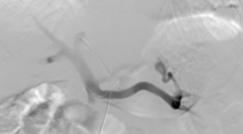
SUMMARY Background: Upper gastrointestinal bleeding from gastroesophageal varices is a frequent complication in patients with liver cirrhosis and portal hypertension. Gastric varices are difficult to control with endoscopic while endovascular intervention is an effective method. We report a case of ruptured gastric varices that was successfully treated by percutaneous transhepatic obliteration. Case presentation : A 50-year-old male patient, with a history of cirrhosis, was admitted to the hospital with a large amount of blood vomiting and black stools. Gastroscopy showed ruptured gastric varices and blood sprayed. The patient was occluded with gastric varices by percutaneous transhepatic obliteration. After 2 days, the patient's condition improved, yellow stools, and was discharged. Conclusion: Obliteration of gastric varices in patients with cirrhosis by percutaneous transhepatic obliteration (PTO) is a safe and effective technique, a good choice in the absence of gastrorenal shunt. The advantage is intervention in emergencies, no need to examine the vessels first with computed tomography. Keywords: Gstric varices, percutaneous transhepatic obliteration.
NGHIÊN CỨU GIÁ TRỊ CỦA CỘNG HƯỞNG TỪ TƯỚI MÁU TRONG CHẨN ĐOÁN VÀ PHÂN LOẠI MÔ BỆNH HỌC UNG THƯ VÚ
12/10/2023 12:44:10 | 0 binh luận
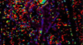
SUMMARY Objective: To investigate wherethe the perfusion parameters of dynamic contrast enhanced magnetic resonance imaging (DCE-MRI) had correlation with molecular subtypes of breast invase ductal carcinoma (IDC). Methods : Forty patients underwent 3T MRI examination following biopsied pathological immunohistochemistry at Bach Mai Hospital from March 2021 to July 2022 were retrospectively investigated. The volume transfer constant (Ktrans), the rate constant (Kep) and the plasma volume ratio (Ve) were calculated from DCE-MRI base on the extended Tofts model. The mean and standard deviation of each perfusion parameters and the pathological immunohistochemistry, subtypes correlations were assessed by Mann-Whitney U test and One-way ANOVA. Results : 40 IDC patients (mean age, 55,5 ± 11.6; range, 38 to 82 years) were investigated. The mean of Kep was higher in tumors with estrogen receptor (ER) negative (p = 0.03), progesterone receptor (PR) negative (p = 0.03), human epidermal growth factor receptor 2 (HER-2) positive (p = 0.02) than that in tumors with ER-positive, PR-positive, and HER-2-negative. Also, Kep was significantly higher in HER-2 enriched tumors than that in luminal B tumors (p = 0.01) and triple-negative tumors (p < 0.05). Conclusion: DCE-MRI may be used as the non-invase tool to assess the molecular biological expression and the molecular subtypes of invase ductal breast cancer. Keywords: Dynamic contrast enhancement, magnetic resonance imaging, subtypes breast cancer, quantitative value.
Bạn Đọc Quan tâm
Sự kiện sắp diễn ra

THÔNG BÁO SINH HOẠT KHOA HỌC 19/01/2024: CẬP NHẬT CHẨN ĐOÁN VÀ ĐIỀU TRỊ UNG THƯ TRỰC TRÀNG
19-01-2024 09:47:01 0 bình luận

Thông tin đào tạo
- Những cạm bẫy trong CĐHA vú và vai trò của trí tuệ nhân tạo
- Hội thảo trực tuyến "Cắt lớp vi tính đếm Photon: từ lý thuyết tới thực tiễn lâm sàng”
- CHƯƠNG TRÌNH ĐÀO TẠO LIÊN TỤC VỀ HÌNH ẢNH HỌC THẦN KINH: BÀI 3: U não trong trục
- Danh sách học viên đạt chứng chỉ CME khóa học "Cập nhật RSNA 2021: Công nghệ mới trong Kỷ nguyên mới"
- Danh sách học viên đạt chứng chỉ CME khóa học "Đánh giá chức năng thất phải trên siêu âm đánh dấu mô cơ tim"


Đơn vị hợp tác

