


NGHIÊN CỨU GIÁ TRỊ CỦA SIÊU ÂM ĐÀN HỒI MÔ NÉN TRONG CHẨN ĐOÁN HẠCH NÁCH ÁC TÍNH
13/10/2023 12:33:32 | 0 binh luận
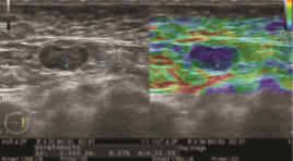
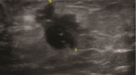
SUMMARY Purpose: To evaluate the accuracy of strain elastography in the diagnosis of malignant axillary lymph nodes. Methods: B-mode and strain elastography US were scanned by experienced radiologists on patients with clinical symptoms or suspicion of breast cancer to look for masses or abnormal lymph node. Lymph nodes are evaluated on B-mode ultrasound based on 4 factors: short and long axis diameter, hilum, and cortical thickening. The strain elastography was based on a color scale of 1 to 5 points based on percentage of nodal region on the color chart, and a semi-quantitative B/A index of node stiffness correlation with adipose tissue of the same depth. Results: a total of 41 lympho nodes (benign, n=13, malignant, n=28) from 39 patients were analyzed. B-mode ultrasonography in the evaluation of malignancy had a sensitivity and specificity of 67.9% and 69.2%. Strain elastography for assessment of axillary lymph node involvement had a sensitivity and specificity of 85.7% and 69.2%. The semi-quantitative index B/A had statistically significant difference between the group of benign and malignant lymph nodes (p<0.05) with the cut of is 23.5. Conclusion: Strain elastography is a non-invasive and helpful method, together with B-mode ultrasound, in the evaluation of malignant axillary lymph node involvement. K ey words: axillary lymph node, strain elastography
NGHIÊN CỨU ĐẶC ĐIỂM HÌNH ẢNH SIÊU ÂM NỘI SOI Ở BỆNH NHÂN UNG THƯ THỰC QUẢN
13/10/2023 12:07:09 | 0 binh luận
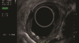
SUMMARY Purpose: The aim of study was decribed the imaging characteristics of endoscopic ultrasound in esophageal cancer. Methods and materials: A cross-sectional descriptive study on 40 patients with esophageal cancer were assigned to endoscopic ultrasound. Results: All study subjects were male with mean age 58.33 ± 7.24. 97.5% of SCC; 2.5% were high-grade dysplasia, none of the patients had adenocarcinoma. Moderately differentiated dominate 55%, 25% highly differentiated and 20% poorly differentiated. .Location of lesions on endoscopy is common in the middle and lower thirds of the esophagus. 85% of esophageal cancer lesions are hypoechoic, 7.5% hyperechoic, and 7.5% mixed. Of which, 70% are heterogeneous, the remaining are homogenous. The lesion size is less than half the circumference accounting for 62.5% and larger than half the circumference accounting for 32.5%. TNM classification on endoscopic ultrasound according to AJCC 8th stage T: Tis (7.5%), T1a (45%), T1b (30), T2 (10%), T3(7.5%). Stage N: N0 (70%), N1 (27.5%), N2 (2.5%). TNM stage: Stage 0 (5%), IA (15%), IB (47.5%), IIB (25%), IIIA (5%), IIIB (2.5%). Conclusion: Endoscopic ultrasound plays an important role in diagnosis and staging in patients with esophageal cancer Key words: endoscopic ultrasound, esophageal cancer, squamous cell carcinoma (SCC), adenocarcinoma.
NHẬN XÉT ĐẶC ĐIỂM LÂM SÀNG VÀ HÌNH ẢNH CẮT LỚP VI TÍNH PHỔI Ở BỆNH NHÂN SAU NHIỄM SARS-COV-2 TẠI CÁC CƠ SỞ KHÁM CHỮA BỆNH CỦA MEDLATEC
13/10/2023 12:01:05 | 0 binh luận
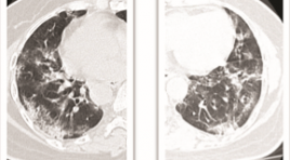
SUMMARY A cross-sectional descriptive study was conducted on 1436 patients after SARS-CoV-2 infection who visited MEDLATEC medical facilities from March 1, 2022 to March 31, 2022. The results showed that 39% were male and 61% female, with a mean age of 39 and the majority of patients in the working age range from 18-60 years old. 74% of patients infected with SARS-CoV-2 have clinical symptoms, of which cough is the most common symptom, followed by chest pain and shortness of breath. The group of patients with a healthy history accounted for 76% of cases; the remaining group of underlying diseases with hypertension accounted for the most, followed by the group with chronic lung disease. Up to 49% of patients have suspected COVID-related lung lesion; with the most important lesions being interstitial thickening and most of them being mild according to the CT-score scale. Keywords: clinical characteristics, CT scanner, SARS-CoV-2
GIÁ TRỊ DỰ BÁO TỬ VONG CỦA ĐỘ NẶNG TỔN THƯƠNG PHỔI TRÊN X-QUANG TẠI THỜI ĐIỂM NHẬP VIỆN Ở BỆNH NHÂN COVID-19
13/10/2023 11:44:33 | 0 binh luận
SUMMARY Background: We aimed to investigate the performance of a chest X-ray (CXR) scoring scale of lung injury in prediction of death among patients with COVID-19 admitted at Vinmec Central Park hospital (HCM City, VN) during the peak epidemic in 2021. Method: Retrospective design; X-ray images and clinical data were collected from all hospitalized patients with SARS-CoV-2 PCR positive from July to September 2021. Three radiologists independently assessed the CXR score at admission which consists of measuring both severity and extent of lung injuries on four lung quadrants (scale o to 24). Association between CXR and mortality risk was estimated using a survival regression with log-log distribution. Result: The study included 219 patients (mortality rate = 28). There was a high consensus for CXR scoring among 3 radiologists (κ = 0.90; CI95%: 0.89-0.92). PCA analysis revealed that CXR has a similar role as CRP score for predicting risk of mortality. CXR score was the strongest predictor of mortality (tdAUC 0.85 CI95% 0.69–1) within the first 3 weeks after admission, compared with other conventional clinical features. After adjusting for the patient’s age, there was a significant effect of increased CXR score on mortality risk (HR = 1.15, CI95%:1.04-1.27, p=0.009). At a critical threshold of 16 points, the CXR score allows for predicting inhospital mortality with good sensitivity (0.82; CI95%: 0.78 to 0.87) and specificity (0.89; CI95%: 0.88 to 0.90). Conclusion: The day-one CXR score is an independent and effective predictor of the risk of death in COVID-19 and could be used to identify high-risk patients in countries like Vietnam where CXR is more readily available than CT scans. Keywords: COVID-19, Chest X-ray, Lung injury score, Mortality prediction
ĐÁNH GIÁ HÌNH THÁI TIỂU NHĨ TRÁI TRÊN CẮT LỚP VI TÍNH ĐA DÃY Ở BỆNH NHÂN TRIỆT ĐỐT RUNG NHĨ QUA ỐNG THÔNG
13/10/2023 11:29:50 | 0 binh luận
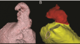
SUMMARY Purpose: to assess left atrial appendage morphology by multidetector computed tomography in atrial fibrillation (AF) patients for catheter ablation Materials and Methods: This retrospective and cross-sectional study included 45 patients diagnosed with AF and treated by catheter ablation and 45 patients without AF undergoing multi-detector computed tomography in E hospital from 1/2020 to 7/2022 Results: The mean age 56.16 ± 11.83, median of left atrium volume in the AF group was 118.13 (96 - 145.56) ml > the control group 72.88 (60.53 - 95.74) ml, (p < 0.001). The most common left atrium morphology in both groups is "cactus" with 46.67% in the AF group and 33.33% in the control group, the middle left atrial appendage orifice was the most common in the AF group (53,33%), while in the control group, the lower left atrial appendage orifice was the most common (44.44%), the length of the left atrial appendage in the AF group was 43.15 ± 8.11 mm, the control group was 44.29 ± 9.76 mm. There is a positive correlation with the mean level between left atrial appendage length and left atrial volume (r = 0.573, p < 0.001). Conclusion: The most common left atrium morphology in both groups is "cactus". There is a positive correlation with the mean level between left atrial appendage length and left atrial volume. Keywords: atrial fibrillation, catheter ablation, left atrial, left atrial appendage, multi-detector computed tomography
ĐÁNH GIÁ ĐẶC ĐIỂM HÌNH ẢNH CỦA CẦU CƠ ĐỘNG MẠCH VÀNH BẰNG CHỤP CLVT 256 DÃY
13/10/2023 11:00:20 | 0 binh luận
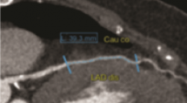
SUMMARY Purpose: to evaluate and analyze the ratio of position, depth and length of the coronary myocardial bridge, the status of pre-myocardial atherosclerosis and the degree of vascular stenosis during systole by multislice-computed tomography (MSCT) images series. Material and method : Computed tomography scan for 175 patients using a 256-row MSCT machine at Hanoi Friendship Hospital between July 2021 and August 2022. Select the best quality images in systole and diastole. Measure the length and thickness of the bridge. For each myocardial bridge segment, assess for the presence of anterior atherosclerotic plaque no more than 2 cm in length of the artery. Assess the degree of vascular stenosis during systole caused by the myocardial bridge. Result: Most of the myocardial bridges were located in the anterior interventricular artery (LAD) (98.3%). The average values of the length and thickness of the bridge are 23.1 ± 10.5mm and 1.0 ± 1.0mm. Atherosclerosis was detected in 77 cases (accounting for 43%). The average degree of tunneling artery stenosis was 23.0% systolic. Conclusion: MSCT 256 series has a high value in detecting bridges, determining the location, there by measuring the length, the thickness of the pons, the status of anterior atherosclerotic plaques as well as the degree of narrowing of the vessel lumen systole of the musculature. Keywords: myocardial bridge, coronary artery, multislice computed tomography.
ĐÁNH GIÁ BƯỚC ĐẦU GIÁ TRỊ CỦA INTERIM PET/CT SAU 2 CHU KỲ ĐIỀU TRỊ HOÁ CHẤT ABVD CỦA BỆNH NHÂN U LYMPHO HODGKIN
13/10/2023 10:44:46 | 0 binh luận
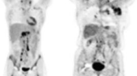
SUMMARY Purpose: The study aimed to initial evaluate the value of interim PET/ CT after 2 cycles of ABVD chemotherapy in Hodgkin lymphoma patients. Material and Methods: The study was carried out retrospective and prospective descriptive study on 56 patients at Tan Trieu K Hospital from March 2020 to June 2022. Patients were examined clinically, paraclinically, had a CT scan or PET/CT film before treatment, then received ABVD chemotherapy, after 2 cycles of being taken and evaluated Interim PET/CT, and continued treatment based on the results. Result of iPET/CT leads to continue or change of regimen continued treatment or change of regimen and follow-up. Results: In a total of 56 patients, male/female ratio: 1/1.6, mean age 30±12.6 (youngest age 7 years old, oldest age 67 years old). 35/56 patients (62.5%) had CT scan and 21/56 patients (37.5%) had background PET/CT scan before treatment. 30.4% had 2 lymph node sites and 28.6 patients had 3 lymph node sites on the body, lymph nodes above the diaphragm 67.9% and lymph nodes both above and below the diaphragm 12,5%. 49 patients had lymph node lesions, short axis size of lymph nodes maximum 27.10±8.3 mm. Lesion to the mediastinum in 15/58 patients (25.8%), spleen in 5 patients. Patients with early stage I-II accounted for 82.1%, advanced stage III-IV accounted for 17.9%. After 2 cycles of ABVD chemotherapy, the response rate on PET/CT was iPET/CT2 (-) 43/56 patients (76.8%, of which Deuville score 1 point 93%) and iPET/CT2 (+) in 13/56 patients (23.3%). The prognostic index of IPS low-risk group (0-2) complete metabolic response was 86.4%. In the high-risk group IPS (4-7) partial metabolic response and complete metabolic response was 54.5% according to Lugano classification. Conclusion: Interim PET/CT has an important role in providing prognostic information in the treatment of ABVD chemotherapy in Hodgkin Lymphoma patients, reducing the toxicity of chemotherapy and determining the next treatment modalities. Keywords: lymphoma Hodgkin, interim PET/CT.
CẬP NHẬN VAI TRÒ CỦA SIÊU ÂM ĐÀN HỒI TRONG CHẨN ĐOÁN UNG THƯ TUYẾN TIỀN LIỆT
12/10/2023 15:57:29 | 0 binh luận
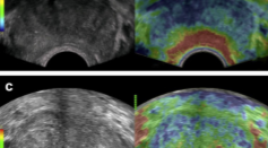
SUMMARY Prostate cancer (PC) is an emerging male health problem in recent years due to new development in diagnostic and treatment modalities that help improve disease prognostic, especially in Eastern countries where PC incidence is high. According to GLOBOCAN 2020, number of new cases of PC reached 1,414,259 while number of deaths was 375,304 cases. In terms of deaths due to cancer in male population, prostate cancer ranks two just after lung cancer. Together with the traditional trio of rectal exam, Prostate-Specific Antigen (PSA) and transrectal ultrasound, emerging imaging modalities such as elastography ultrasound, multimodality magnetic resonance imaging not only helped physicians better detecting malignant lesions at supra early stage but also guided biopsy for pathological assessment. These new innovations led to preference of target-biopsy with lower rate of re-biopsy and complications comparing to systemic biopsy technique
Bạn Đọc Quan tâm
Sự kiện sắp diễn ra

THÔNG BÁO SINH HOẠT KHOA HỌC 19/01/2024: CẬP NHẬT CHẨN ĐOÁN VÀ ĐIỀU TRỊ UNG THƯ TRỰC TRÀNG
19-01-2024 09:47:01 0 bình luận

Thông tin đào tạo
- Những cạm bẫy trong CĐHA vú và vai trò của trí tuệ nhân tạo
- Hội thảo trực tuyến "Cắt lớp vi tính đếm Photon: từ lý thuyết tới thực tiễn lâm sàng”
- CHƯƠNG TRÌNH ĐÀO TẠO LIÊN TỤC VỀ HÌNH ẢNH HỌC THẦN KINH: BÀI 3: U não trong trục
- Danh sách học viên đạt chứng chỉ CME khóa học "Cập nhật RSNA 2021: Công nghệ mới trong Kỷ nguyên mới"
- Danh sách học viên đạt chứng chỉ CME khóa học "Đánh giá chức năng thất phải trên siêu âm đánh dấu mô cơ tim"


Đơn vị hợp tác

