


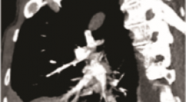
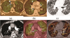
GIẢ PHÌNH ĐỘNG MẠCH PHỔI: NHÂN 3 TRƯỜNG HỢP KHÁM VÀ ĐIỀU TRỊ TẠI BỆNH VIỆN PHỔI TRUNG ƯƠNG
11/10/2023 15:21:47 | 0 binh luận
SUMMARY Pulmonary artery pseudoaneurysms (PAPs) is a rare abnormality of the pulmonary artery system. Beside, PAPs have no specific clinical presentation or may be asymptomatic, which may be present in congenital anomalies or in a variety of conditions such as pneumonia, pulmonary neoplasm, pulmonary tuberculosis, or lung fungus... [1]. We present 3 cases of PAPs examined and treated at the National Lung Hospital with PAPs appearing on the background of 3 different diseases. Both of these 3 cases have nonspecific symtoms as cough, chestpain and dyspnea with 2 cases of hemoptysis. They are all detected by computed tomography (CT) angiography. There are 2 cases treated by surgery that have good results and 1 case who continued the TB regimen received medical treatment. Keywords: pulmonary artery pseudoaneurysms, pulmonary artery, hemoptysis.
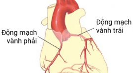
GIÁ TRỊ CỦA CẮT LỚP VI TÍNH 256 DÃY TRONG CHẨN ĐOÁN BỆNH ĐỘNG MẠCH VÀNH Ở BỆNH NHÂN RUNG NHĨ
11/10/2023 15:05:26 | 0 binh luận
SUMMARY Objectives: To determine the diagnostic value of 256 MDCT in the diagnosis of coronary artery disease in patients with atrial fibrillation. Materials and methods: We prospectively enrolled 48 patients with atrial fibrillation who underwent 256 MDCT coronary CT angiography scan at Huu Nghi Hospital from 07/2020 to 07/2021 and evaluated diagnosis value of this technique using invasive coronary angiography as golden standard. Results: At patient-based analysis, sensitivity, specificity, positive predictive value and accuracy are 100%, 83.3% and 83.3%, respectively. At vessel-based analysis, sensitivity, specificity, positive predictive value, negative predictive value and accuracy are 98.4%, 81.7%, 80.3%, 98.5% and 88.9%, respectively. At segment-based analysis sensitivity, specificity, positive predictive value, negative predictive value and accuracy are 96.7%, 93.8%, 78.4%, 99.2 % and 94.4%, respectively. Conclusion: 256 MDCT coronary angiography scan is a diagnostic method with high accuracy in diagnosing coronary artery disease in patients with atrial fibrillation. Keywords: atrial fibrillation, coronary CTA, diagnostic value
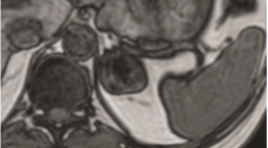
VAI TRÒ CỦA CỘNG HƯỞNG TỪ TRONG CHẨN ĐOÁN U TUYẾN THƯỢNG THẬN
11/10/2023 15:01:16 | 0 binh luận
SAMMARY Purpose: to evaluate the role of MRI in diagnosis adrenal tumors. Subject and methods: our cross–sectional descriptive study included 25 patients who were diagnosed as adenoma hyperplasia in pathology. All patients underwent MRI examination in Bach Mai hospital from June, 2020 to July, 2021. Results: mean age of all patient was 49,2±11,8. Male/female ratio was 1.5/1. 25 adrenal tumor on MRI and identified on the diagnosed as adenoma hyperplasia in pathology. These adenomas consisted 12 adrenal cortical adenoma (ACA) and 13 non – adrenal cortical adenoma (NACA): 09 pheochromocytomas, 01 adrenocortical carcinoma, 01 ganglioneuroma and 02 myelolipoma. Mean size of all adrenal tumors was 38,8 ± 23,5mm, therein, mean size of ACA group and NACA group was 23,2 ± 7,6 mm and 52,2 ± 25,7 mm respectively. Chemical Shift Imaging (CSI) allowed differential diagnose between ACA and NACA. Qualitative analysis based on signal reduction on out – phase compared to in – phase allows 11/12 ACA, but 2/13 NACA have been misdiagnosed as ACA. Quantitative analysis based on SII index (SII threshold > 16,1%) allowed correctly diagnose 11/12 ACA and 01 NACA. ASR index (ASR threshold <0,71) allowed correctly diagnose 11/12 ACA and 01 NACA. There are 02 adrenal tumors that invaded to the surrounding area in imaging and was demonstrated by surgery and pathology reports. Conclusion : MRI has an important role in adrenal mass diagnosis with sensitivity and specificity were 100% and 96,1% respectively. CSI enable to distinguish ACA and NACA with sensitivity and specificity were 91,7% and 92,3% respectively by using SII and ASR index. MRI also help to assess invasive and metastatic properties with high accuracy. Key words : adrenal tumors, adrenal MRI, chemical shift imaging (CSI).
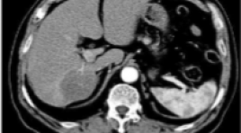
ĐÁNH GIÁ HIỆU QUẢ ĐIỀU TRỊ UNG THƯ BIỂU MÔ TẾ BÀO GAN BẰNG NÚT MẠCH VI CẦU PHÓNG XẠ YTTRIUM-90
11/10/2023 14:52:30 | 0 binh luận
SUMMARY Background: Hepatocellular carcinoma (HCC) is the second most common cause of cancer-related deaths worldwide. Radioembolization with Yttrium-90 is a safe, effective locoregional therapy for unresectable HCC. Purpose: This study aimed to evaluate the safety and efficacy of radioembolization with Yttrium-90 for the treatment of HCC. Materials and methods: A total of 93 patients were diagnosed with HCC and treated by radioembolization with Yttrium-90 from 2013 to 2021 at Bach Mai Hospital. Radiographic findings on computed tomography (CT) or magnetic resonance imaging (MRI) were assessed by using modified Response Evaluation Criteria in Solid Tumors (mRECIST). Patient survival was assessed using the Kaplan-Meier method, and prognostic factors affecting survival were assessed using Cox proportional hazards regression. Results: The mean age of 93 patients was 59.0±9.8 years. Mean tumors diameter was 6.5 ± 2.7 cm, the mean of tumor marker AFP is 1812 ± 6577 ng/ml. Complete response and partial response to treatment were observed in 21 (40.4%) and 15 patients (28.8%) of 52 follow-up patients. The median overall survival (OS) was 12.0 [95% CI: 5.0-25.0] months. Factors associated with longer OS included BCLC stage A-B, ECOG 0 and no portal vein thrombosis. Conclusion: Radioembolization with Y-90 is safe and effective in patients with unresectable HCC. BCLC stage A-B, ECOG 0 and no portal vein thrombosis were positive prognostic factors. Keywords: Hepatocellular carcinoma, radioembolization, Selective internal radiation therapy, Yttrium-90, Tumor response
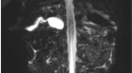
ĐẶC ĐIỂM HÌNH ẢNH CHT 4 CA TEO ĐƯỜNG MẬT BẨM SINH THỂ CÓ NANG GIỐNG U NANG ỐNG MẬT CHỦ
11/10/2023 14:44:26 | 0 binh luận
SUMMARY Purpose: to describle in detail the significant signs in hepatobiliary MRI, that help to give different diagnosis between biliary atresia (BA) with big cyst at hepatic hilar and choledochal cyst in small childrent and infant. Method: presenting 4 cases with clinical diagnosed of biliary atresia that based on clinical signs and US, MRI results, all 4 cases were operated, taken cholangiogram in surgery to confirm the diagnosis of cystic biliary atresia and had pathological results. Results: All 4 patients were under 3 months old, all are female with enlarged liver; gallbladder were in the normal size limit but all of them had abnormal shape: deformation, irregular wall, communicated directly with common bile duct. All 4 cases have cystic diameter over 10mm. No case of all has further biliary signal that above or under the cyst on MRI but one case has contrast material filling intraliver bile duct draining directly to cystic lesion on cholangiogram intraoperation . These are 3 cases of cystic BA type III and 1 cases of cystic BA type I. 2 cases has positive triangular cord sign (TC sign). Conclusion : Hepatobiliary MRI with its basic sequences, specially with MRCP is helpful to give difirential diagnosis of cystic BA and choledochal cyst in the young childrent. Keyword: Biliary atresia, cystic biliary atresia
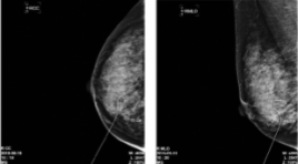
KẾT QUẢ BAN ĐẦU SINH THIẾT VÚ HÚT CHÂN KHÔNG DƯỚI HƯỚNG DẪN SIÊU ÂM CÓ KẾT HỢP ĐỊNH VỊ KIM CHO TỔN THƯƠNG VI VÔI HÓA
11/10/2023 14:38:52 | 0 binh luận
SUMMARY Purpose: This study examined the usefulness of ultrasound-guided vacuum-assisted breast biopsy for mammographic microcalcification. Methods : case series from June- 2019 to June-2020 at Breast department Cho Ray hospital. The patients with BI-RADS Category 4 Mammographic microcalcification were included. Most microcalcifications were not observed on ultrasound. Sono-guided J-wire localization was first performed for the suspicious microcalcification area, and the location of the J-wire and calcification was determined with mammography in most cases. Sono-guided VABB was performed after removing the J-wire without a stereotactic device. On the other hand, Sono-guided VABB was performed directly without J-wire localization when microcalcification lesions were identified by mass on ultrasonography. In all cases, calcification was confirmed by specimen mammography and the pathology was performed. A follow-up examination was performed to confirm the presence of complications. Results: A total of 20 lesions of 18 patients with BI-RADS Category 4 Mammographic microcalcification were included. Mean age: 49,44 ± 9,49 (35-66). Mean size of lesions 10,83 ± 3,60 mm, (4-15mm). In 20 lesions, 6 lesions (30%) were diagnosed as a malignancy (2 cases of ductal carcinoma in situ, 3 cases of ductal carcinoma invasive, 1 case of atypical ductal hyperplasia). The remaining 14 lesions (70%) were diagnosed as benign (fibroadenoma: 4; fibrocystic exchange 7, fibrocystic desease: 1, typical hyperplasia : 2). There were no significant complications during follow up after VABB. Conclusion: Sono-guided VABB can be used effectively if combined with wire localization for microcalcification lesions. Keywords: Mamography, Calcification, VABB, Image-guides biopsy, J Wire localization
ĐẶC ĐIỂM HÌNH ẢNH SIÊU ÂM VÀ CẮT LỚP VI TÍNH TRONG CHẨN ĐOÁN VIÊM TÚI THỪA MANH TRÀNG
11/10/2023 14:31:15 | 0 binh luận
SUMMARY Objectives: Describe the characteristics of ultrasound images and computed tomography in the diagnosis of cecal diverticulitis. Subjects and methods : A cross-sectional study on 49 patients who underwent ultrasound and computed tomography at the Center of Radiology in Bach Mai Hospital, diagnosed with cecal diverticulitis on CT, was treated at Bach Mai Hospital from June 2020 to June 2021. Results: ultrasound showed diverticulum 63.3%, inside diverticulum there was fecal stone 18.3%, gas or fecal content 18.3%, fluid 26.5%, diverticulum wall thickness (>2mm) 28.6%, cecal wall thickness 95.9%, fat infiltration around the diverticulum or caecum 81.6%. 47/49 patients had thickened cecal wall, 89.8% thickened around the circumference. CT scan, 71.4% had 1 diverticulum, 20.4% had multiple diverticula, 10.2% had gas in the diverticulum, 55.1% had fecal stones, 26.5% had fluid in the diverticulum, thickened diverticulum (>2mm) 71.4%, 95.9% thickening of the cecum wall. Conclusions : Ultrasonography and CT imaging are very valuable imaging tools for determining the diagnosis, extent of lesions and complications of cecal diverticulitis, and are meaningful in clinical practice for choosing treatment methods. Keywords: cecal diverticulitis, ultrasound, computed tomography
CHỤP CẮT LỚP VI TÍNH HAI MỨC NĂNG LƯỢNG PHÁT HIỆN TẮC ĐM PHỔI: LỢI ÍCH THÊM VÀO CỦA BẢN ĐỒ IODINE
11/10/2023 12:57:49 | 0 binh luận
Summary Purpose: To determine if there is an added benefit of using iodine maps from dual-energy (DECT) in addition to conventional CT angiography images to diagnose pulmonary embolism (PE). Materials and Methods : In this retrospective analysis, 49 consecutive dual-energy CT angiography examinations performed from August through July 2020 at Bach Mai Hospital to evaluate for PE were reviewed. The 49 examinations included 49 patients (mean age, 59.73 years; range, 22–99 years). First, the location, level, and type (occlusive vs nonocclusive) of PEs on conventional CT angiograms were recorded. Iodine maps were then reviewed for defects suggestive of PE. Last, CT angiograms were rereviewed to detect additional PEs suggested by the iodine map. Results: 19/49 (38.8%) patients were diagnosed with PE, a total of 247 PEs were detected at initial review. After review of the DECT iodine map, 16 additional PEs were found on 8 of 49 (16.3%) patients in which 2 of 49 (4 %) patients had a new diagnosis of PE after review of the DECT iodine maps, 4/49 (8%) patients were diagnosed PE before. Of the 16 additional PEs, 8 (50%) were segmental, 8 (50%) were subsegmental, 3 (18.8 %) were occlusive, and 13 (81.2%) were nonocclusive Conclusion: Dual-energy CT iodine maps show a small incremental benefit for the detection of occlusive segmental and subsegmental pulmonary emboli. Keywords: CTPA= computed tomography pulmonary angiography, DECT= dual-energy computed tomography, PE= pulmonary embolism, iodine map.
Bạn Đọc Quan tâm
Sự kiện sắp diễn ra

THÔNG BÁO SINH HOẠT KHOA HỌC 19/01/2024: CẬP NHẬT CHẨN ĐOÁN VÀ ĐIỀU TRỊ UNG THƯ TRỰC TRÀNG
19-01-2024 09:47:01 0 bình luận

Thông tin đào tạo
- Những cạm bẫy trong CĐHA vú và vai trò của trí tuệ nhân tạo
- Hội thảo trực tuyến "Cắt lớp vi tính đếm Photon: từ lý thuyết tới thực tiễn lâm sàng”
- CHƯƠNG TRÌNH ĐÀO TẠO LIÊN TỤC VỀ HÌNH ẢNH HỌC THẦN KINH: BÀI 3: U não trong trục
- Danh sách học viên đạt chứng chỉ CME khóa học "Cập nhật RSNA 2021: Công nghệ mới trong Kỷ nguyên mới"
- Danh sách học viên đạt chứng chỉ CME khóa học "Đánh giá chức năng thất phải trên siêu âm đánh dấu mô cơ tim"


Đơn vị hợp tác

