


KHẢO SÁT VỊ TRÍ LỖ THÔNG XOANG BƯỚM Ở NGƯỜI TRƯỞNG THÀNH TRÊN CẮT LỚP VI TÍNH
17/10/2023 15:27:45 | 0 binh luận
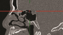
SUMMARY Background: The sphenoid sinus is closely surrounded by many important vascular and neurological. The natural ostium is the safest place to enter the sphenoid sinus without injuring adjacent structures. A better understanding of the position and distance of the sphenoid ostium (SO) with respect to the other anatomic landmark is essential for the safety and effectiveness of laparoscopic surgery. Objective: The purpose of this study was to investigate the relationship between the SO and surrounding landmark structures. Besides, we examine the effect of the Onodi cell and pneumatization of the sphenoidal sinus on the position of the SO. Methods: Cross-sectional study of 162 sinus CT scan data obtained from the PACS system at University Medical Center Ho Chi Minh City. The image is reconstructed and measured by the Multiplanar reconstruction (MPR) software of the PACS Carestream. Results: The mean distance between the SO and the lateral wall was 9.1 ± 1.8 mm. The mean distance from the SO to the median line was 4.1 ± 1.8 mm. The mean distance between the SO and the roof sphenoid sinus was 7.9 ± 2.9 mm. The average distance from the sphenoid ostium to superior border of posterior choana was 12.9 ± 3.5 mm. The mean distance from the sphenoid ostium to the anterior nasal spine was 65.2 ± 4.5 mm and to the posterior wall of the sphenoid sinus was 12.8 ± 2.7 mm. The angle between the SO - the anterior nasal spine and the floor of the nose is 33.5 ± 3.5 degrees. The distance from the SO to the roof sphenoid sinus was found to increase in cases where Onodi cells are present. For lateral pneumatization, the distance from the SO to the lateral wall in type II to be less in type III. The sphenoid sinus pneumatization on the sagittal plane did not affect the distance from SO to the posterior choana and the roof sphenoid sinus. Conclusions: The study determined the distances between the SO and some surrounding anatomical landmarks, and also investigated the influence of the Onodi cell and pneumatization on the position of the sphenoid sinus by CT scan images. The measurements described in this study may be very valuable in avoiding serious complications while performing surgery. Keywords: Sphenoid ostium, computed tomography.
ĐẶC ĐIỂM HÌNH ẢNH SIÊU ÂM, CỘNG HƯỞNG TỪ VÀ CHỤP MẠCH SỐ HOÁ XOÁ NỀN CỦA MỘT SỐ BỆNH NHÂN TĂNG SẢN LÀNH TÍNH TUYẾN TIỀN LIỆT
17/10/2023 15:05:17 | 0 binh luận
SUMMARY Objective: Describe Imaging features Ultrasound, MRI and DSA of 14 patients who have benign prostatic hyperplasia with acute urinary retention, were treated by PAE. Method and results: A descriptive study without control in patients hospitalized for acute urinary retention was diagnosed with benign prostatic hyperplasia at the Bach Mai hospital from 07/2017 to 07/2018. The patients were examined clinical, ultrasound, MRI before and after intervention. The average PV (cm3) on ultrasound and MRI are 80,57±50,58 cm3 and 82,35±51.24 cm3, 9 patients have thickening of bladder wall (64,3%), 3 patients have bladder diverticulum (21,4%), 78,6% hyperplasia at the transition zone, 7,1% hyperplasia at the central zone and 14,3% hyperplasia at both the transition zone and central zone. On DSA, with 26 pelvices side were perfomed successful, there were 22 pelvices side with one prostate artery and 4 pelvices side with 2 prostate arteries, the average diameter of prostate with pelvic side which one prostate artery is 1,92 ± 0,52 mm; with pelvic side which two prostate arteries is 1,45 ± 0,16mm. Keywords : prostatic arterial embolization, acute urinary retention, benign prostatic hyperplasia, ultrasound, MRI of BPH.
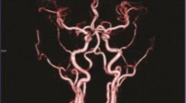
NGHIÊN CỨU TƯƠNG QUAN GIỮA CHỤP CẮT LỚP VI TÍNH 640 LÁT CẮT VÀ CHỤP MẠCH SỐ HÓA XÓA NỀN TRONG CHẨN ĐOÁN BỆNH LÝ ĐỘNG MẠCH VÀNH TẠI BỆNH VIỆN ĐA KHOA QUỐC TẾ VINMEC ĐÀ NẴNG
17/10/2023 12:18:28 | 0 binh luận
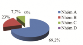
SUMMARY Purposes: To descriptive coronary artery imaging by the 640 slice coronary computed tomography scanning. To determine the value between coronary computed tomography scanning angiography and digital subtraction angiography. Materials and methods: Describe imaging characteristics and stratify. There are 310 patients with the 640 slice coronary computed tomography scanning Toshiba Aquillion One in the period from 1.1.2019 to 30.8.2021 and among whom 42 cases followed by digital subtraction angiography (DSA). Results: The average age is 59,5±12,5 years old and male/female about 3/1. HR 68,28±18,67 bpm. Radiation dose < 5 mSV (90%), average dose 3,79±0,96mSv. Calcium score is mostly mild level, distributed all arterial branches. LAD is accounted for the highest percentage of calcification(> 50%) and many narrow position. The most narrow position is proximal location and the most narrow level is mild. The value table Value Se (%) Sp (%) Accuracy (%) LM 100 100 100 LAD 97,5 100 97,6 LCx 90,9 90,0 95,2 RCA 93,3 91,7 92,9 Conclusion: .Coronary computed tomography scanning evaluated anatomy, abnormal orginal position, variant and the narrow of position and level. Digital subtraction angiography has difficultly identifying abnormal variant except myocardial brigde. The values with sensitivity, specificity and accurary of coronary artery stenosis assessement by computed tomography scanning angiography showed high. Keywords: coronary aretry, coronary computed tomography scanning, digital subtraction angiography, coronary artery anatomy, coronary artery variant.
GIÁ TRỊ KẾT HỢP CỦA SIÊU ÂM ĐÀN HỒI SÓNG BIẾN DẠNG VÀ CHỌC HÚT TẾ BÀO BẰNG KIM NHỎ TRONG CHẨN ĐOÁN UNG THƯ VÚ
17/10/2023 12:02:16 | 0 binh luận
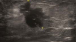
Objective: Describe characteristics of 2D ultrasound images and shear wave elastography (SWE) ultrasound breast tumor. Determinate the combined value of SWE ultrasound and fine needle aspiraton cytology (FNAC) in diagnosis breast cancer. Subject and method: 79 female patients with breast tumors were assigned 2D and SWE ultrasound, measure shear wave velocity (SWV) internal tumor and peripheral tumor, classified according to ACR - BIRADS 2013. Patients were assigned FNAC ultrasound guided and histopathological biosy after surgery. Compare the result 2D, SWE ultrasound and FNAC with the result of histopathological biopsy after surgery to determine the combined value of SWE ultrasound and FNAC in diagnosis breast cancer. Result: 79 patients with 37 benign breast tumors, 42 malignant breast tumors. Mean SWV of malignant breast tumors are lower than benign breast tumors (p < 0.01). Cutoff SWV in the differential diagnosis of benign and malignant breast tumor at peripheral tumor 2.25 m/s; internal tumor 5.1 m/s. SWE ultrasound combined with FNAC has high value in diagnosis breast cancer with Se = 97.6%, Sp = 94.6%, Acc = 96.2%, PPV = 95.3%, NPV = 97.2%, high concordance with histopathological result after surgery Kappa = 0.924. Keywords : Elastography, fine needle aspiraton cytology, cancer, breast.
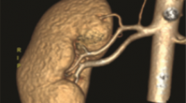
NGHIÊN CỨU GIÁ TRỊ CỦA CẮT LỚP VI TÍNH 256 DÃY TRONG ĐÁNH GIÁ GIẢI PHẪU MẠCH MÁU THẬN ĐOẠN NGOÀI THẬN Ở NGƯỜI SỐNG HIẾN THẬN
17/10/2023 11:45:34 | 0 binh luận
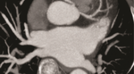
SUMMARY Objective: To determine the accuracy of 256-slice computed tomographic angiographyin the evaluation of renal arterial and venous structures of living donor kidney system and its anatomical variations in living kidney donors, and the correlation of CTRA findings with those observed during kidney harvesting Materials and Methods: 202 potential living donor candidates were included to this study, who had CTA for the assessment of their renal vessels in our hospital between January 2021 and June 2022. The number, course, and drainage patterns of the renal vessel were retrospectively observed from the scans. Anomalies of renal arteries, veins and inferior vena cava (IVC) were recorded. Multiplanar reformations (MPRs), maximum intensity projections, and volume rendering were used for analysis. The results obtained were correlated surgically. Results: Two-hundred and two patients who had undergone laparoscopic nephrectomy as living donors were included: 109 female and 93 male donors Mean age of donors was 33.19± 7.09 years. Bilateral renal arteries mostly originate at the level of the L1 vertebral body and the L1/2 disc. Mean diameter of right renal artery was 5.94 ± 0.94mm and left renal artery was 5.90±0.94mm. The mean length of the right renal artery was 33.9 ± 13.5 mm and left renal artery was 27.8 ± 10.1 mm. The distribution of accessory renal artery was 15.3% on right side and 19.8% on left side. 8.9% has multiple renal veins, more common in the right side. Sn, Sp, NPV, PPV compare open oparative nephrectomy 93.75-100%. Conclusion: 256-slice computed tomographic is an accurate, safe and noninvasive diagnostic tool for determination of renal artery and vein abnormalities in living kidney donors preooparative. It helps with surgery planning, choosing operation side and exclusion of donors. Keywords: Kidney transplantation; Living renal donor; Computed tomography angiography; Renal artery

ĐÁNH GIÁ KẾT QUẢ CHỌC HÚT MÁU TỤ NỘI SỌ TRÊN LỀU TỰ PHÁT DƯỚI ĐỊNH VỊ KHÔNG KHUNG VÀ CẮT LỚP VI TÍNH
17/10/2023 11:21:32 | 0 binh luận
SUMMARY Objectives: To evaluate the results of spontaneous intracranial hematoma aspiration under frameless positioning and computed tomography. Methods: Retrospective study of 55 patients (patients) diagnosed with spontaneous intracranial hematoma (MTNS) by computed tomography (CT) scan from 05/2017 to 04/2022 at 108 Central Military Hospital. Results: The mean volume fraction of residual hematoma was 26.24% after draining for an average of 2-3 days, respectively. There was no significant difference in the percentage of residual hematoma volume after draining. The favorable 1-month outcome with GOS 4 or 5 was significantly better in the group with a hematoma reduction of more than 60% compared with baseline hematoma volume (p=0.047), although no significant difference was observed. told at 6 months after aspiration. The factor that was significantly correlated with favorable outcome after aspiration 6 months was the ratio of final hematoma volume after drainage (p=0.016). Final hematoma volume ≤ 15 ml was correlated with favorable neurological outcomes at 1 and 6 months (p = 0.001 and 0.038). Conclusion: There was no difference in the final residual hematoma volume and neurological outcome after 6 months depending on the time of aspiration aspiration of the hematoma. The factor influencing neurological outcome after 6 months of aspiration is the final hematoma volume remaining after drainage. Final hematoma volume ≤15ml was correlated with favorable neurological outcome at 1 and 6 months. This study provides the goal for hematoma aspiration technique to leave a residual hematoma volume ≤ 15ml. Keywords: Spontaneous supratentorial intracerebral hematoma, computed tomography, frameless navigation
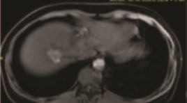
GIÁ TRỊ CỘNG HƯỞNG TỪ XÓA NỀN 3.0 TESLA ĐÁNH GIÁ TĂNG SINH MẠCH Ở BỆNH NHÂN UNG THƯ BIỂU MÔ TẾ BÀO GAN SAU TACE
17/10/2023 11:01:54 | 0 binh luận
SUMMARY Objective: The value of dynamic subtraction MRI 3.0 Tesla technique in the angiogenesis assessment of hepatocellular carcinoma after transcatheter arterial chemoembolization. Methods: A cross-sectional, retrospective descriptive study was performed on 40 patients, who had been diagnosed with HCC and treated with TACE, then do dynamic subtraction MRI 3T from 6/2021 to 6/2022 at Bach Mai hospital, Hanoi, Vietnam. Result: The study consisted of 40 patients with 57 hepatocellular carcinoma lesions who underwent transarterial chemoembolization procedure and followed up by dynamic MRI of the liver with post processing to obtain subtraction images and compared with DSA. The subtraction images have sensitivity of 100%, specificity of 100%, PPV of 100%, and NPV of 100%. On the other hand, the dynamic images is 90,9%; 69,2%; 90,9%;69,2%; the Diffusion images is 97,7%; 61,5%; 89,6%; 88,9%. Comparative study between D-MRI and DS-MRI assessments revealed; highly significant increase in disease detection rate, sensitivity, and NPV in favor of DS-MRI in HCC patients; with highly significant difference (p < 0.01 respectively). Conclusions: Dynamic MRI is valuable in the angiogenesis assessment of hepatocellular carcinoma after TACE, however, this value is augmented by the addition of subtraction technique especially in lesions having high signal before administration of contrast medium with sensitivity of 100%, specificity of 100%, PPV of 100%, and NPV of 100%. So we recommend adding the subtraction technique in the protocol of MRI in the follow up after transarterial chemoembolization as it increases the diagnostic confidence. Keywords: dynamic subtraction MRI, hepatocellular carcinoma, TACE.
XÁC ĐỊNH TỔN THƯƠNG ĐƯỜNG DẪN TRUYỀN GIỮA HAI BÁN CẦU ĐẠI NÃO SAU CHẤN THƯƠNG ĐẦU BẰNG HÌNH ẢNH HỌC MRI - DTI (DTI: DIFFUSION TENSOR IMAGING): BÁO CÁO CA LÂM SÀNG
16/10/2023 17:13:10 | 0 binh luận
SUMMARY Traumatic brain injury (TBI) is a form of acquired brain injury resulting from an external mechanical force to the head make up the alternation of brain function. However, it is difficult to diagnose the injury to the neural tract and the connection between us through traditional imaging techniques. A 54y woman came to our clinic because of insufficient coordination of her body. Her personal history: Severe trauma brain injury with coma in 10 days treated by medical treatment 10 years ago. Some bad conditions after TBI include impairment of memory, insufficiency of coordination of her body, a post-concussion syndrome was suggested. Clinical examination: GCS: 15, strength testing: 5/5 for both sides but our patient can’t walk. She was examined at many medical centers but no evidence conforming her symptom. Our patient scanned by MRI ((Magnetic resonance imaging) Siemens 3.0 Tesla Spectra system. MRI morphometry used for her detected a significant decline of corpus callosum. MRI - DTI (Diffusion Tensor Imaging) revealed a decreased FA in the white matter of the right temporal and corpus callosum. Fractional anisotropy is a scalar value between zero and one that describes the degree of anisotropy of a diffusion process. A decrease in the value of FA in corpus callosum is indicative of the loss of connection between both hemispheres. MRI tractography used to describe the amount of neural tracts in corpus callosum. So, MRI-DTI and MRI Tractography served as a powerful diagnostic tool, providing imaging results that offered an explanation for our patient’s clinical picture Keyword: Trauma brain injury, Cerebral atrophy, Diffusion tensor imaging, Tractography, Morphometry
Bạn Đọc Quan tâm
Sự kiện sắp diễn ra

THÔNG BÁO SINH HOẠT KHOA HỌC 19/01/2024: CẬP NHẬT CHẨN ĐOÁN VÀ ĐIỀU TRỊ UNG THƯ TRỰC TRÀNG
19-01-2024 09:47:01 0 bình luận

Thông tin đào tạo
- Những cạm bẫy trong CĐHA vú và vai trò của trí tuệ nhân tạo
- Hội thảo trực tuyến "Cắt lớp vi tính đếm Photon: từ lý thuyết tới thực tiễn lâm sàng”
- CHƯƠNG TRÌNH ĐÀO TẠO LIÊN TỤC VỀ HÌNH ẢNH HỌC THẦN KINH: BÀI 3: U não trong trục
- Danh sách học viên đạt chứng chỉ CME khóa học "Cập nhật RSNA 2021: Công nghệ mới trong Kỷ nguyên mới"
- Danh sách học viên đạt chứng chỉ CME khóa học "Đánh giá chức năng thất phải trên siêu âm đánh dấu mô cơ tim"


Đơn vị hợp tác

