


NGHIÊN CỨU GIÁ TRỊ CỦA KỸ THUẬT CHỌC HÚT TẾ BÀO BẰNG KIM NHỎ DƯỚI HƯỚNG DẪN CỦA SIÊU ÂM TRONG CHẨN ĐOÁN HẠCH DI CĂN Ở BỆNH NHÂN UNG THƯ TUYẾN GIÁP THỂ BIỆT HÓA
16/10/2023 17:08:53 | 0 binh luận
SUMMARY Background: Ultrasonic-guided fine needle aspiration cytology is a minimally invasive diagnostic technique with the highest accuracy and sensitivity in the diagnosis of lymph node metastasis. Objectives: The study consists of 2 objectives: 1) Describe clinical and subclinical characteristics in treated patients with differentiated thyroid cancer; Evaluation of the role of ultrasound-guided fine needle aspiration cytology in the diagnosis of lymph node metastasis in patients with differentiated thyroid cancer. Materials and methods: A cross-sectional descriptive study design was conducted on 89 patients diagnosed with differentiated thyroid cancer and found that the cervical lymph nodes were potentially malignant under ultrasonography and indicated fine-needle aspiration cytology under ultrasound guidance. Results: Among 89 research subjects, small cell aspiration of lymph node groups, group IV and VI lymph nodes with suspicion of malignancy accounted for the highest proportion of 25/89 nodes (28.1%) fine needle aspiration cytology, in which positive accounts for 30.1%. Sensitivity, specificity, negative predictive value, false positive rate, false negative rate, and cytology accuracy were 90%; 100%; 100%; 52.9%.0%, 10.1%, 91.01%. Conclusion: Ultrasonic-guided fine needle aspiration cytology for the diagnosis of lymph node metastasis in differentiated thyroid cancer has a low false-positive rate, high accuracy, accurately reflecting the presence or absence of underlying cervical lymph node metastasis. Avoid complications and thoroughly treat metastatic disease for patients. Keywords: Ultrasonic-guided fine needle aspiration cytology, metastatic lymph nodes, differentiated thyroid cancers
NGHIÊN CỨU MỐI LIÊN QUAN GIỮA NỒNG ĐỘ THYROGLOBULIN, THỜI GIAN NHÂN ĐÔI THYROGLOBULIN VỚI TỔN THƯƠNG TÁI PHÁT, DI CĂN TRÊN HÌNH ẢNH 18F-FDG PET/CT Ở BỆNH NHÂN UNG THƯ TUYẾN GIÁP THỂ BIỆT HÓA KHÁNG 131I
16/10/2023 17:03:32 | 0 binh luận
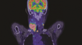
SUMMARY Background: 18F-FDG PET/CT is method with high sensitivity and specificity in the diagnosis of recurrent lesions in patients with radioiodine refractory differentiated thyroid cancer ((RAI-R- DTC). The aim of this study was to evaluate the relationship between Tg, Tg – DT and rucurrent/metastatic lessions detected in 18F-FDG PET/CT imaging in RAI-R DTC patients. Methods: 119 RAI-R DTC patients underwent 18F-FDG PET/CT. At least two consecutive Tg measurements under the thyroid hormone replacement therapy (TSH ≤ 0,1 uIU/ml) to calculate Tg - DT before 18F-FDG PET/CT scan. We analyzed the relationship between PET/CT imaging and clinical characteristics, risk of recurrence, Tg and Tg – DT. Results : 18F-FDG PET/CT imaging detected recurrent lesions in 88 patients (73,9 %) and distant metastasis in 29 patient (24,3 %). Tg-DT was significantly lower in the positive PET/CT group compared to negative PET/CT group (median 12.9 vs 166 months, p< 0.001). Serum Tg concentration in the patients with distant metastasis was higher than those without distant metastasis (334 ng/ml vs 114.6 ng/ml, p < 0.001). In univariate and multivariate logistic regression analysis, Tg-DT was an independent predictor of positive PET/CT and serum Tg concentration was a predictor of distant metastasis. The ROC curve determines the optimal threshold of Tg - DT in predicting positive PET/CT is 34.75 months and Tg level at 218.1 ng/ml predicting distant metastasis Conclusion: Tg - DT is reliable value in predicting positive 18F - FDG PET/CT and serum Tg concentration is valuable for predicting the outcome of distant metastasis in RAI – R DTC patients. Keywords : thyroglobulin, thyroglobulin doubling time, differentiated thyroid carcinoma, 18F - FDG PET/CT.
TÍNH TOÁN CHE CHẮN CHO PHÒNG XẠ TRỊ GAMMA DÙNG PHẦN MỀM MONTE CARLO CODE EGSNRC
16/10/2023 16:54:04 | 0 binh luận
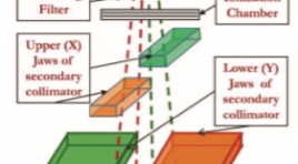
SUMMARY This study applies the Monte Carlo simulation method EGSnrc Code with two dedicated codes: BEAMnrc code is used to simulate the beam emitted from the accelerator head and DOSXYZnrc code is used to calculate the dose emitted from the accelerator. From there, evaluate the beam attenuation of radiation emitted from the accelerator through the layers of shielding material. Initial study results showed that dose limited at staff area (area Control) is 0,11 mSv/ week (5,5 mSv / year) and in the public area (area Uncontrol) is 0,022 mSv/week (1,1 mSv/year). Initial research results show that the effectiveness of the application of the EGSnrc simulation program with two specialized codes, BEAMnrc and DOSXYZnrc, shows that the radiation dose in the study area for medical staff is safe, within the allowable limits. Keywords: Accelerator, Radiation Safety, MCNP, EGSnrc, DOSXYZnrc, and BEAMnrc
NGHIÊN CỨU ĐẶC ĐIỂM TỔN THƯƠNG XƯƠNG TRÊN XẠ HÌNH Ở BỆNH NHÂN UNG THƯ PHỔI KHÔNG TẾ BÀO NHỎ GIAI ĐOẠN IIB - IVB TẠI BỆNH VIỆN UNG BƯỚU THÀNH PHỐ CẦN THƠ NĂM 2020 - 2021
16/10/2023 16:48:04 | 0 binh luận
SUMMARY Background: Non-small cell lung cancer (NSCLC) is the most common cancer and has a high rate of bone metastases. Early detection of bone metastases is important in treatment and improves the quality of life for. Currently, there have been many studies on the value of bone scan in early detection of bone metastases, but this issue has not been fully evaluated. Objective: To evaluate the characteristics of bone metastases on scintigraphy in lung cancer. Methods: Cross-sectional description of a series of diseases, retrospective data on 151 patients with stage IIB - IVB non-small cell lung cancer, examined by bone scan with Tc-99m MDP (Methylene diphosphonate) before specialized treatment during the period from January 2020 to December 2021 at Can Tho City Oncology Hospital. Results: The incidence of men and women were 62,9% and 37,1%. The mean age is 60, the youngest is 35 and the oldest is 83. There are 56 patients (37,1%) with bone lesions on the scan. There are 47 lesions (83,9%) could be known as bone metastases: 46 cases are at stage IV and the other is at stage IIIB. The most common sites of lesions are from the ribs and sternum. Others are from thoracic spine, the sacrum and the coccyx. The bone lesions from the clavicle and upper extremities are rare. Most of bone lesions are multifocal, asymmetrical, and strongly radioabsorbed. Conclusion: Bone-image characteristics from scan in stage IIB – IVB non-small cell lung cancer have a high incidence of bone metastases. Therefore, the application of bone scan as a routine subclinical in the initial diagnosis of NSCLC before treatment is essential in order to properly assess the stage of the disease, make an accurate prognosis and have a reasonable treatment strategy. Keywords: Bone scan, non-small cell lung cancer
ĐẶC ĐIỂM HÌNH ẢNH SIÊU ÂM CỦA HẠCH VÙNG CỔ Ở BỆNH NHÂN UNG THƯ TUYẾN GIÁP THỂ BIỆT HÓA ĐÃ ĐIỀU TRỊ
16/10/2023 16:33:28 | 0 binh luận
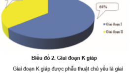
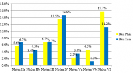
SUMMARY Background: Ultrasound is a minimally invasive diagnostic technique with the highest accuracy and sensitivity in the diagnosis of lymph node metastasis. Objectives: The study consists of objectives: Describe the ultrasound imaging characteristics of cervical lymph nodes in treated patients with differentiated thyroid cancer. Materials and methods: A cross-sectional descriptive study design was conducted on 89 patients diagnosed with differentiated thyroid cancer and found that the cervical lymph nodes were potentially malignant under ultrasonography and indicated fine-needle aspiration cytology under ultrasound guidance. Results: Among 89 study subjects, the main reason for admission was lymphadenopathy, accounting for 95.5%. The results of papillary pathology accounted for 96.6%. Time after thyroid surgery when cervical lymph node metastasis is detected is 1-3 years (27%). The time after thyroid surgery when cervical lymph node metastasis was detected was 6-36 months (38.2%). Oval shape on ultrasound has 73%, cortex on ultrasound accounts for 65.2%, decreased lymph node on ultrasound accounts for 64%, loss of hilar lymph nodes on ultrasound accounts for 62.9%. Conclusion: Ultrasound plays an important role in diagnosing the stage of the disease, fully and accurately describing the lesions and stage of the disease, helping to increase the effectiveness of treatment. Keywords: ultrasonic imaging, lymph nodes, differentiated thyroid cancers.
ĐÁNH GIÁ SỰ CẢI THIỆN MỨC ĐỘ TRƯỢT THÂN ĐỐT SỐNG SAU PHẪU THUẬT TLIF DỰA TRÊN X-QUANG THƯỜNG QUY
16/10/2023 16:25:53 | 0 binh luận
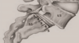
SUMMARY Aim: The study was carried out with the aim of evaluating the improvement of degree of vertebral body slippage after spinal fixation posterior slide manipulation with disc removal, by combining vertebaral body fusion TLIF surgery and assessingthe improvement in the level of post-X-ray-based vertebral slip, with clinical comparison. Method: A total of 39 patients diagnosed with lumbar vertebral stem slide who were surgically treated at the Department of Orthopedic and Spinal Injuries of Bach Mai Hospital between July 2021 and July 2022 were included in the study. Results: The results of the study showed that patients with lumbar vertebral slippage experienced the most in the L4-L5 position accounting for 67.4%, followed by L3-L4 accounting for 22.5%, L5-S1 accounting for 13.1%. All 100% of patients improved in the height of the disc gap between the vertebrae. No more diseases showed signs of ladder. Signs of intermittent pain also improved, and 80 percent of patients were able to travel longer distances than before. Conclusion: Postoperative X-ray examination showed that thepatients were corrected in surgery quite well, all patients had reduced levels of postoperative slippage and increase intervertebral space height. Keywords: routine X-ray, lumbar vertebral stem slide, TLIF surgery
ĐẶC ĐIỂM HÌNH ẢNH CỘNG HƯỞNG TỪ 3.0 TESLA CỦA U NGUYÊN BÀO VÕNG MẠC
14/10/2023 12:30:07 | 0 binh luận
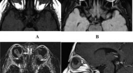
SUMMARY Purpose: To describe the imaging characterization of high-resolution magnetic resonance imaging of retinoblastoma Material and method: About 50 patients were diagnosed and monitored for retinoblastoma treatment at the National Eye Hospital and performed 3T magnetic resonance at Saint Paul General Hospital from October 2020 to May 2022. Description of tumor intraocular lesion: Number, location of tumor, largest diameter. Description of tumor characteristics: Signals on sequences T1W, T2W, FLAIR, DWI and enhancement properties after injection. Other accompanying features: Intra-tumor calcification, retinal detachment, vitreous hemorrhage, optic nerve invasion, invasion outside the eyeball, intracranial lesions Results: 50 patients in the study had an average age of 21.2 months, and the age of the one-eye disease group was higher than that of the two-eyed disease group. 3T MRI is a good method for diagnosing retinoblastoma tumors, with the largest dimension measuring 13.8 ± 4.2 mm, the tumor distribution is mainly on the retina in the post-equatorial region. Tumor image on MRI compared with vitreous signal, mostly increased signal on T1W (92.6%), decreased on T2W (98.8%), increased on FLAIR (100%), restriction on DWI (100%) and enhancement after injection. Signs of calcification are specific for retinoblastoma, however, on MRI the ability to detect calcification is limited. Although the detection of calcification is limited, it has good ability in invasive assessment such as choroidal invasion (15.6%), scleral invasion (3.1%), optic nerve invasion (31.3). %). Severe prognostic signs such as retinal detachment (34.4%), vitreous bleeding (26.6%). Conclusion: 3T magnetic resonance is a good method of diagnosing retinoblastomas as well as assessing the invasiveness of tumors. Keywords: Retinoblastoma, High resolution magnetic resonance imaging of retinoblastoma
NGHIÊN CỨU KẾT QUẢ BƯỚC ĐẦU ĐIỀU TRỊ UNG THƯ BIỂU MÔ TẾ BÀO GAN BẰNG PHƯƠNG PHÁP ĐỐT SÓNG CAO TẦN TRONG MỔ
14/10/2023 12:09:21 | 0 binh luận
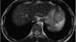
SUMMARY Intraoperative radiofrequency ablation is a new approach for the radical treatment of patients with multifocal hepatocellular carcinoma in different lobes. Originally, patients no longer indicated for radical treatment were now surgically removing the tumor in one lobe and radiofrequency ablation of other tumors in the surgery period time. Objectives and subjects: A retrospective and prospective study conducted between 2020 and 2022, 09 patients with HCC with at least 2 tumors in different lobes underwent intraoperative radiofrequency ablation (IORFA) and followed-up for 3 months at Bach Mai Hospital. Results: All 09 patients with 28 tumors, the number tumors from 02 to 06, each patient underwent surgery to remove 01 large tumor and radiofrequency ablation in surgery for other tumors. All patients responded completely according to mRECIST criteria, no local recurrence, relapsed or died within the first 3 months. Conclusion: Intraoperative radiofrequency ablation is a new effective method approach, help to expand the indications for radical treatment for patients with multifocal hepatocellular carcinoma. Keywords: Intraoperative radiofrequency ablation, hepatocellular carcinoma. *
Bạn Đọc Quan tâm
Sự kiện sắp diễn ra

THÔNG BÁO SINH HOẠT KHOA HỌC 19/01/2024: CẬP NHẬT CHẨN ĐOÁN VÀ ĐIỀU TRỊ UNG THƯ TRỰC TRÀNG
19-01-2024 09:47:01 0 bình luận

Thông tin đào tạo
- Những cạm bẫy trong CĐHA vú và vai trò của trí tuệ nhân tạo
- Hội thảo trực tuyến "Cắt lớp vi tính đếm Photon: từ lý thuyết tới thực tiễn lâm sàng”
- CHƯƠNG TRÌNH ĐÀO TẠO LIÊN TỤC VỀ HÌNH ẢNH HỌC THẦN KINH: BÀI 3: U não trong trục
- Danh sách học viên đạt chứng chỉ CME khóa học "Cập nhật RSNA 2021: Công nghệ mới trong Kỷ nguyên mới"
- Danh sách học viên đạt chứng chỉ CME khóa học "Đánh giá chức năng thất phải trên siêu âm đánh dấu mô cơ tim"


Đơn vị hợp tác

