


NGHIÊN CỨU ĐẶC ĐIỂM HÌNH ẢNH 18FDG-PET/CT HẠCH CỔ Ở BỆNH NHÂN UNG THƯ VÒM MŨI HỌNG
14/10/2023 11:57:56 | 0 binh luận
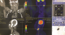
SUMMARY Purpose: Describe imaging characteristics and evaluate the role of 18FDG-PET/CT in the diagnosis of cervical lymph nodes in nasopharyngeal cancer patients. Subjects and methods: Prospective description of 60 patients with nasopharyngeal carcinoma identified by biopsy and histopathology and untreated (in which 55 patients with undifferentiated carcinoma and 5 patients with squamous cell carcinoma), were taken with 18FDG PET/CT and compared with ultrasound and done FNA lymph nodes respectively, from June 2018 to July 2019 to K Central Hospital, Tan Trieu campus. All 18FDG-PET/CT films were read and compared with their respective locations on ultrasound and lymph node FNA before treatment. Results: In total 60 patients common age is 52.3±11.1, male/female ≈ 5/1; A total of 379 bilateral cervical lymph nodes on PET/CT, 195 lymph nodes in the left neck are common (51.5%), the most common lymph node location in group II (96.7% of patients and 61% of total lymph nodes). There are 322/379 (85%) lymph nodes with structural loss on ultrasound, there is a correlation between lymph node size and lymph node absorption on 18FDG-PET/CT with r=0.6, cervical lymph node groups 322/379 Structural abnormal lymph nodes with SUVmax = 9.5 ± 4.6 (nearly 4 times higher than 57/379 normal lymph nodes with SUVmax = 2.6 ± 2.5). In a total of 379 lymph nodes over 60 patients, there was a close agreement between the lymph nodes with loss of umbilical fat structure on ultrasound with SUVmax>2.5 threshold of 92.9% (kappa=0.67) and better at the threshold. SUVmax>3.5 was 92.9% (Cohen's kappa=0.75) on PET/CT. In addition, cytology was positive in 60/89 lymph nodes undergoing FNA, which was in close concordance with ultrasonography of loss of hilar fat structure and cytological diagnosis of 83.4% (Cohen's kappa = 0.62). ). The cytological diagnosis was mild with increased uptake with SUVmax>2.5 threshold of 75.3% (Cohen's kappa = 0.3), but there was a strong correlation when the lymph node imaging was increased. Absorption with SUVmax >2.5 on PET/CT and loss of umbilical cord fat on ultrasound with cytological diagnosis was 87.6% (Cohen's kappa = 0.69). In particular, out of a total of 340/379 lymph nodes with threshold SUVmax>2.5 on PET/CT are considered as metastatic nodes, changing the stage of 25/60 patients (41.67%); in which 17 patients increased and 8 patients decreased stage N compared with staging by other imaging methods before having PET/CT. Conclusion: 18FDG-PET/CT scan is an imaging method with accurate diagnostic value in diagnosing cervical lymph nodes in patients with cervical cancer, useful for treatment planning. Key words: lymph nodes, nasopharyngeal cancer, 18FDG-PET/CT lymph nodes on nasopharngeal cancer.
ĐẶC ĐIỂM HÌNH ẢNH PHÌNH ĐỘNG MẠCH NÃO TRÊN CỘNG HƯỞNG TỪ 3 TESLA
13/10/2023 18:04:10 | 0 binh luận
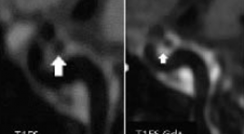
SUMMARY Objective: Describe the imaging characteristics of an intracranial aneurysm on 3 Tesla magnetic resonance at Bach Mai hospital Methods : A prospective study was performed on 29 patients with clinical manifestations of neuropathy who were diagnosed with 3 Tesla magnetic resonance brain aneurysms and then underwent digital subtraction angiography at the Radiology Center, Bach Mai Hospital, Hanoi, Vietnam from August 2021 to August 2022. Description of intracranial aneurysms in terms of number, location, shape, morphology and size measure. Results: Of the 29 patients, 37 aneurysms were detected on 3 tesla magnetic resonance and confirmed on digital subtraction angiography. The location of intracranial aneurysms is common in the anterior circulation (97.29%), and the posterior circulation (2.71%). Dimensions are mainly ≤ 10mm (97.3%). Usually saccular aneurysms (94.6%) and narrow neck (51.43%). Most aneurysms have irregular margins, knobs or two bases (72.97%). Conclusion : Magnetic resonance 3 Tesla can replace digital subtraction angiography erasing the background to characterize the image of intracranial aneurysms. This is a safe, non-invasive and highly valuable imaging method in the diagnosis of intracranial aneurysms, a very effective first choice for screening intracranial aneurysms and providing information for neurologists implement appropriate treatment strategies. Keywords: 3T magnetic resonance, intracranial aneurysm
GIÁ TRỊ CỦA CỘNG HƯỞNG TỪ ĐỘNG SÀN CHẬU TRONG CHẨN ĐOÁN HỘI CHỨNG ĐẠI TIỆN TẮC NGHẼN
13/10/2023 17:58:31 | 0 binh luận
SUMMARY Objectives : to describe the clinical features and images of MR defecography in patients with obstructive defecation syndrome, afterward evaluating the value of MR defecography in the diagnosis of obstructive defecation syndrome. Subjects and methods: cross-sectional study, 33 patients were diagnosed with obstructive defecation syndrome according to ROME IV criteria, were assigned to have MR defecography at Saint Paul General Hospital, during the period of study from January 2019 to July 2022. Results: About the general characteristics of the study group: mainly in women with 84,8%, the average age is 63.5 ± 12.5 years old. Rectocele is the most commonly detected lesion on clinical examination, with 12/33 cases, accounting for 36.4%. About image characteristics on pelvic floor dynamic MRI: the mean value of H line in normal phase is 4.7 ± 0.9cm, in strain phase is 5.9 ± 1.5cm. The mean value of the M-line in normal phase is 2.0 ± 0.2 cm, and 4.1 ± 0.3 cm in strain phase. Rectocele is the most common lesion, accounting for 63.6%. The detection rate of rectocele and associated lesions (cystocele, uterus descent, cervical descent, vaginal descent) on MR defecography is higher than that on clinical examination, the difference was statistically significant with p<0.05. Conclusion : Our study contributes to clarifying the high applicability of MR defecography in diagnosing the causes and grading of pelvic floor prolapse, pelvic organ prolapse in patients with obstructive defecation syndrome, combined with clinical symptoms to help clinicians make appropriate treatment indications for each patient. Key words: MR defecography, Obstructed defecation syndrome, Rectocele. *
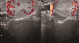
ĐÁNH GIÁ HIỆU QUẢ ĐIỀU TRỊ U LÀNH TUYẾN GIÁP CÓ ĐIỂM THẨM MỸ ≥ 3 BẰNG ĐỐT SÓNG CAO TẦN
13/10/2023 17:49:10 | 0 binh luận
SUMMARY Purpose: To evaluate the efficacy of radiofrequency ablation (RFA) for benign thyroid neoplasms with a cosmetic score of more than 3. Materials and Methods: This prospective study was performed on 60 patients with benign thyroid nodules with a cosmetic score of more than 3 by radiofrequency ablation under ultrasound guidance at 108 Military Central Hospital from January 1. 2021 to July 2022. We evaluated the change in cosmetic, volume, angiogenesis, and clinical symptoms of patients at 1, 3, and 6 months after the intervention as well as complications, complications during intervention and treatment monitoring. Results : Most patients are female, accounting for 91.7%. The most common age group is from 30 to 50 years old, accounting for 45% and the average age is 43.2 years old. Grade 3 and grade 4 cosmetic scores are equivalent. After 6 months of RFA, the angiogenesis score decreased to 24%, followed by the volume of thyroid nodules decreasing to 37%. The score of symptoms and cosmetics decreased by about 50% compared to before treatment. Complications were mild pain and mild hematoma after RFA, accounting for 5%. Conclusion: Radiofrequency ablation is an effective and safe method for improving the cosmetic point of the neck in the treatment of benign thyroid nodules. Keywords: Benign thyroid nodules, cosmetic score ≥ 3, radiofrequency ablation.
ĐÁNH GIÁ KẾT QUẢ ĐIỀU TRỊ ĐAU DÂY V BẰNG PHƯƠNG PHÁP PHẪU THUẬT GIẢI ÉP VI MẠCH VÀ TIÊM CỒN KHOANG MECKEL
13/10/2023 17:31:51 | 0 binh luận
SUMMARY Objective: To evaluate the results of treatment of trigeminal neuralgia by microvascular decompression surgery and Meckel’s cavity alcohol injection Subject and method: 62 patients with trigeminal neuralgia which medical treatment did not control the pain or had side effects, the patient then received microvascular decompression surgery or Meckel’s cavity alcohol injection performed at Saint Paul general Hospital from January 2017 to June 2021 Results: From January 2017 to June 2021, 62 patients with trigeminal neuralgia underwent microvascular decompression surgery or Meckel’s cavity alcohol injection at Saint Paul general Hospital. Of these, 24 patients were treated by surgical decompression and 38 patients were treated with Meckel’s cavity alcohol injection. The results of pain relief time of both methods in both short and long term are not statistically significant with p<0.05 according to Log rank test. The complication rate of the two methods is similar right after the intervention but will be lower in the surgical group after 3 months of intervention (p < 0.05 in Chi-squared test). Conclusion: The short and long-term analgesic effect of the two methods of microvascular decompression surgery and Meckel’s cavity alcohol injection are similar, the difference is not statistically significant. Key words: Microvascular decompression surgery, Meckel’s cave alcohol injection, Trigeminal neuralgia.
GIÁ TRỊ CỦA CẮT LỚP VI TÍNH TRONG ĐÁNH GIÁ DỊ VẬT ỐNG TIÊU HÓA SẮC NHỌN
13/10/2023 17:05:44 | 0 binh luận
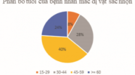
SUMMARY Ingested foreign bodies are one of the reasons why patients need to be hospitalized in the emergency room. Although most gastrointestinal foreign bodies can be eliminated from the body on their own, there are still some cases that cause complications, even death. In terms of foreign body morphology, there are 3 main groups, sharp-pointed foreign bodies, round foreign bodies and long foreign bodies, of which sharp-pointed foreign bodies are the group that often causes complications, especially perforation complications. There are many methods to diagnose this condition such as X-ray, computed tomography (CT) and endoscopy. In recent years, the popularity of CT has made this method widely used in the assessment of sharp-pointed gastrointestinal foreign bodies. Objective: In order to evaluate the effectiveness of CT in diagnosing sharp foreign bodies, we would like to conduct the study "Value of computed tomography in evaluation of sharp-pointed ingested foreign bodies". Subjects and research methods: A descriptive study of 25 patients with sharp-pointed foreign bodies in the gastrointestinal tract who underwent CT scan at the Radiology Department of Hanoi Medical University Hospital for 2 years, and underwent endoscopic and surgical or percutaneous intervention to remove of foreign bodies. Results: The most common site for foreign bodies was the jejunum, followed by the stomach, colon, and esophagus. Transmural foreign body accounted for the highest rate (14/25 cases). The most common complication of sharp-pointed foreign body on CT is perforation with 16/25 cases, followed by abscess with 04/25 cases. CT scan has high sensitivity and specificity in evaluating complications of sharp-pointed foreign bodies. Conclusion: CT has an important role in detecting and diagnosing sharp-pointed gastrointestinal foreign bodies as well as evaluating associated complications. Key word: Ingested foreign bodies, foreign bodies, complications of foreign bodies, sharp-pointed foreign bodies, sharp-pointed
ĐẶC ĐIỂM HÌNH ẢNH CLVT ĐA DÃY VỠ XƯƠNG HÀM MẶT DO TAI NẠN GIAO THÔNG
13/10/2023 16:55:55 | 0 binh luận
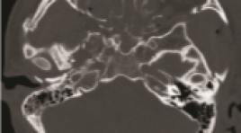
SUMMARY Objectives : study the multi-slice CT (MSCT) imaging in the diagnosis of maxillofacial fractures by traffic accident. Subjects and methods: A cross-sectional descriptive study of 63 patients with maxillofacial trauma due to traffic accidents who underwent MSCT scan at Viet Duc Hospital in April 2022. Results: 46 male and 17 female. The mean age was 28.4±12.6 (from 15 to 75 years old). The most common traffic accidents are motorbikemotorcycle accidents with 21/63 (33.3%), self-motorbike-riding accidents are 18/63 (28.6%), car-motorcycle accidents account for 16 /63 (25.4%), pedestrian - car/motorcycle accidents are 5/63 (7.9%) and car-car accidents are 3/63 (4.8%). There were 28/63 (44.4%) cases associated with cranial fracture and 32/63 (50.8%) skull base fracture. In cases of maxillofacial fracture, orbital wall fracture was the most common with 41/63 (65.1%), maxillary fracture 39/63 (61.9%), zygomatic fracture 32/63 (50.8%), nose bone 19/63 (30.2%), frontal sinus wall 13/63 (20.6%), palatal and mandibular fracture were 11/6 (17.5%). The most common type of maxillary fractures is Lefort 1 with 11/63 (17, 5%) on both right and left side, Lefort 2 and 3 fractures are less common with 1.6% to 6.3%, respectively. Conclusion : MSCT is a reliable method in diagnosing maxillofacial fractures. Keywords: maxillofacial trauma, traffic accident, computer tomography.
NHẬN XÉT SỰ AN TOÀN CỦA DẪN LƯU BỂ THẬN QUA DA DƯỚI HƯỚNG DẪN CỦA SIÊU ÂM VÀ DSA Ở BỆNH NHÂN TẮC NGHẼN ĐƯỜNG BÀI XUẤT CAO
13/10/2023 16:35:42 | 0 binh luận
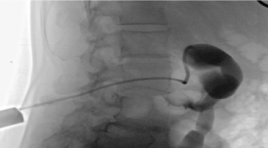
SUMMAR Aim : to access the safety and complications of Percutaneous Nesphrostomy in obstructed urinary upper tract. Patients and methods : a cross-sectional descriptive study was performed on 45 patients who were diagnosed with pyelonephritis and hydronephrosis due to obstructed urinary upper tract at the Radiology Department, Ha Noi Medical Hospital from 6/2021 to 8/2022. Results: 100% of patients who have hydronephrosis and pyelonephritis taken broad-spectrum antibiotics before performing percutaneous nephrostomy (PCN). Position to drain into renal pelvis: 93.3% from the inferior pole, 6.7% from the middle pole. 91.1% of patients were performed successfully Percutaneous Nephrostomy the first time and 8.9% of patients needed in the second time. The successful rate of Percutaneous Nephrostomy was 100%. Time to complete procedure on average 19.5±4.5 minutes. Bleeding complications accounted for 2.2% (n=1), Sepsis complications accounted 6.7% (n=3). Catheter obstruction was 18% (n=8). The size of the sonde drainage 8F was used for patients with Hydronephrosis and pyelonephritis in grades I and II. The size of the sonde drainage 10-12F was used for patients with Hydronephrosis and Pyelonephritis in grades III and IV. Conclusion: Percutaneous nephrostomy is a minimal, safe, effective treatment procedure. Keywords : Renal Nephrosis, Pyelonephritis, Percutaneous Nephrostomy
Bạn Đọc Quan tâm
Sự kiện sắp diễn ra

THÔNG BÁO SINH HOẠT KHOA HỌC 19/01/2024: CẬP NHẬT CHẨN ĐOÁN VÀ ĐIỀU TRỊ UNG THƯ TRỰC TRÀNG
19-01-2024 09:47:01 0 bình luận

Thông tin đào tạo
- Những cạm bẫy trong CĐHA vú và vai trò của trí tuệ nhân tạo
- Hội thảo trực tuyến "Cắt lớp vi tính đếm Photon: từ lý thuyết tới thực tiễn lâm sàng”
- CHƯƠNG TRÌNH ĐÀO TẠO LIÊN TỤC VỀ HÌNH ẢNH HỌC THẦN KINH: BÀI 3: U não trong trục
- Danh sách học viên đạt chứng chỉ CME khóa học "Cập nhật RSNA 2021: Công nghệ mới trong Kỷ nguyên mới"
- Danh sách học viên đạt chứng chỉ CME khóa học "Đánh giá chức năng thất phải trên siêu âm đánh dấu mô cơ tim"


Đơn vị hợp tác

